Quantcast
Channel: Life is short, but snakes are long
Viewing all 105 articles
Browse latest View live

Linnaean Snakes: Part I

$
0
0
This article will soon be available in Spanish
Este artículo pronto estará disponible en español

Although recent findings have shed new light on the (so far) oldest-known fossil snakes, extending the fossil record of snakes back in time an incredible 70 million years, this article is about a more anthropocentric definition of "the first snakes". It's about the first snakes to be named and described using the modern system of classification: those described and classified by Linnaeus in the 10th edition of his Systema Naturae, using consistently together for the first time a binomial naming system for genera and species and a hierarchical category system for higher taxa (i.e., families, orders, classes, phyla, and kingdoms). Although Darwin's theory of evolution has ultimately refocused modern taxonomy on cladistics and phylogenetic trees, the Linnaean system is not wholly incompatible with our new understanding of the common ancestry of all life, and has and will continue to be used.

Carl Linnaeus (left) and Peter Artedi (right)
Carl Linnaeus was primarily a botanist, coining Latin names for and describing over 7,700 species of plants in his lifetime. However, he did a pretty good job of naming and describing species of animals as well, with over 4,400 to his name. His interest in describing animals derived partly from an agreement he made with his friend and one-time rival, Peter Artedi, when the two men were students: that if either of them should die, the other would complete their life's work. Artedi, an ichthyologist, drowned at age 30 (wrote Linnaeus, "too early...did the most distinguished of ichthyologists perish in the waters, having devoted his life to the discovery of their inhabitants!"), so Linnaeus took it upon himself to organize, complete, and publish Artedi's work on the classification of fishes. In truth, the two men developed the basics of zoological nomenclature together, and if Artedi had lived he probably would have shared equally in the renown which has come to Linnaeus today.

Tantilla melanocephala from the King of Sweden's collection
The snakes that Linnaeus described came primarily from a few sources. Several small collections ('curiosity cabinets') made by European aristocrats and businessmen formed the basis of a handful of his zoological dissertations, short papers written primarily by Linnaeus and defended by his students at the University of Uppsala, as was the custom at the time. One such dissertation, Amphibia Gyllenborgiana (defended by B. R. Hast in 1745), describes a collection donated by the university chancellor, Count Carl Gyllenborg, which contained the first attempt to classify snakes according to their numbers of scales, rather than their colors or patterns. Another, Surinamensa Grilliana (defended by Peter Sundius in 1748), describes a collection acquired with the help of Claes Grill, a wealthy merchant with an interest in natural history who used his directorship of the Swedish East India Company to obtain plants and animals from Surinam. Some of these specimens are still in the museum in Uppsala, including a caecilian, two Red Pipesnakes, a false coralsnake, and a parrotsnake. These dissertations do not use the binomial nomenclature for which Linnaeus is now famous. A few years later, Linnaeus was asked by the King and Queen of Sweden to organize, describe, and publish accounts of their personal natural history collections. In those days, it was as fashionable to collect objects of natural history, such as shells, insects, and preserved specimens, as it is to collect art today. The king in particular had amassed a large collection of snakes, many of which are still in the Swedish Museum of Natural History today (and looking remarkably well for being almost 300 years old), and these are described in Linnaeus's 1754 Museum Adolphi Friderici. During the 1750s and 60s, many of Linnaeus's students (which he called his "apostles") traveled the world collecting and sending him specimens, but in accordance with his interests they mostly sent him plants. A few students, including Pehr Kalm, who explored and collected in North America, and Fredrik Hasselqvist, who explored the Middle East, sent Linnaeus a few reptiles. Almost half of the snakes in Systema Naturae are from the king's collection, and most of the others are from the collections and works of two Dutch naturalists whose collections Linnaeus had seen as a young traveler: Albertus Seba, who wrote a Thesaurus of animals with many engravings (including hundreds of snakes), and Laurens Theodorus Gronovius, who worked mostly on fish (the distinction between fish and reptiles was still a bit hazy at the time). Although Linnaeus no doubt could have read about other snakes, he was skeptical of anything he had not examined himself1, and limited his published descriptions to specimens he could examine personally.

Title page of the 10th edition
In the 10th edition of his Systema Naturae, Linnaeus listed a total of 110 species in the order Serpentes, in six genera: Crotalus, Boa, Coluber, Anguis, Amphisbaena, and Caecilia. The first three will be familiar to any snake enthusiast, but the latter three, while legless, have since been reclassified as lizards or amphibians2. Of the 100 species that are actually snakes, 74 are still considered valid today. Linnaeus added 18 more snake species in his 1766 12th edition3, 13 of which are still valid, for a grand total of 87 snake species currently bearing his name, over 2% of modern species; only the authors of Erpétologie Générale can claim more. For reptiles as a whole he still ranks as the 9th most prolific taxonomist4. Pretty good for a botanist. To be fair, Linnaeus had the distinct advantage of Systema Naturae's 10th edition being later declared the starting point of zoological nomenclature, so he has benefited from having any names which preceded his automatically invalidated, whatever their notoriety. His cavalier attitude towards the work of those who came before him rankled many of his contemporaries, although he cited their descriptions wherever he could verify them. This also means that it was technically impossible for him to have "redescribed" any taxa, as many later authors often did, even though in reality of course many kinds of snakes were already recognized and some had names dating back to antiquity (many of which he used). Only 14 of the 100 snake species in SN10 were described therein for the first time. All these advantages didn't stop him from naming invalid species though—26 of the 100 species in the 10th edition (and 5 in the 12th) he described twice, under two different names; that is, later herpetologists decided that the specimens in his descriptions were members of the same species and synonymized (or "lumped") them, which accounts for the reduction in his total number of snake species from 118 to 87.

The travels of Linnaeus's students (click for larger version)
Linnaeus worked on classifying many different groups of organisms, and he always worked in great haste, because there was so much to do. As a result, he could be fairly careless, particularly when it came to the geography of his specimens (i.e., his type localities). Because he had not actually been to many of the places where his specimens came from, he had to rely on the word of others for this information. When specimens came from his apostles or from other contemporaries, they usually had pretty accurate, if general, locations (e.g.,'America', 'Africa'). If they were older, such as those in the collections of Gronovius, Seba, and the king, they were often accompanied by unverifiable locations, many of which were incorrect. In fact, only 33 of the 74 snake species in Linnaeus's SNX have unambiguously correct location information. A further 21 are unambiguously wrong, and 20 bear the label 'Indiis', which might refer either to India or to the West Indies (and, in either case, is still incorrect for certain specimens). In certain cases, it almost seems that labels were switched, such as a South American Xenodon from 'Asia' and an Asian Amphiesma from 'America'. Overall his snakes are fairly diverse, with good geographic representation, except for Australia, which was first botanized in 1770, close to Linnaeus's death, by Linnaean apostle Daniel Solander, sailing onboard James Cook's Endeavour along with Joseph Banks (and resulting in the name of Botany Bay).

Many other later taxonomists reorganized Linnaeus's snake genera, breaking up his combinations by placing the vast majority of the snakes Linnaeus described into new genera. However, 4 of his snake species retain their original genus and species names today. That three of them would was inevitable because of the principle of priority and the "type" concept5, but the fourth is a bit of a bonus. Next month, in Part II, we'll take a closer look at these four species, named by Linnaeus when George Washington was in his twenties, 257 years ago.


1 Seba's Thesaurus contained a now-famous image of a hydra, which Linnaeus inspected in Hamburg in 1735 and exposed as a hoax made from weasels and snake skins. This and other mythical creatures he listed as "animalia paradoxa" in early editions of Systema Naturae, although some (like the paradoxical frog) turned out to be real! For instance, he was correct in stating that "All the other dragons listed by authors are fictitious, like the hydra, which I saw at Hamburg, but which was an outstanding work, not of nature, but of art.", but erred in thinking that "The horned viper is a coluber fabricated by the craft of the Arabs, who pierced its head with the claws of a small bird and then inserted them there".


2 Originally, two scolecophidians (Amerotyphlops reticulatus and Typhlops lumbricalis), the monotypic Anilius scytale, a pipesnake (Cylindrophis maculatus), and two sand boas (Eryx colubrinus and E. jaculus) were placed in Linnaeus's genus Anguis, but were later reclassified (correctly) as snakes.


3 Nothing new was added to the 11th edition, which was simply a reprint of the 10th. In 1789, 13 years after Linnaeus's death, Johann Friedrich Gmelin added three more species of snakes to the 13th and last edition, by which time other zoologists such as Laurenti (who also split reptiles from amphibians and tripled the number of reptile genera) had already contributed a great deal to snake taxonomy.


4 It's fair to say that Linnaeus didn't like snakes or other reptiles. In the first edition of Systema Naturae he wrote: "The Creator in his benignity has not wanted to continue any further the Class of Amphibia for, if it should enjoy itself in as many Genera as the other Classes of Animals, or if those things were true that the Tetralogists have fabricated about Dragons, Basilisks, and such monsters, the human genus would hardly be able to inhabit the earth."He continues in Museum Adolphi Friderici: "Truly formidable are the arms which the Lord of nature has given to some animals. Though he has left serpents destitute of feet, wings, and fins, like naked fishes, and has ordered them to crawl on the ground exposed to all kinds of injuries, yet he has armed them with dreadful envenomed weapons: but, that they may not do immoderate mischief, he has only given these arms to about a tenth part of the various species; at the same time arraying them in such habits that they are not easily distinguishable from one another, as the rest of animals are; so that men and other creatures, while they cannot well distinguish the noxious ones from those which are innocent, shun them all with equal care. We shudder with horror when we think of these cruel weapons. Whoever is wounded by the Hooded Serpent (Coluber Naja) expires in a few minutes; nor can he escape with life who is bitten by the Rattle-snake (Crotalus horridus) in any part near a great vein. But the merciful God has distinguished these pests by peculiar signs, and has created them most inveterate enemies; for as he has appointed cats to destroy mice, so has he provided the Ichneumon [mongoose] (Viverra Ichneumon) against the former serpent, and the Hog to persecute the latter. He has moreover given the Crotalus a very slow motion, and has annexed a kind of rattle to its tail, by the motion of which it gives notice of its approach...On account of these and various other poisonous serpents and worms of India, which crawl upon the ground, swim in the waters, or twine among the branches of trees, we prefer our barren and craggy woods to the everblooming meadows and fruitful groves of Indian climes; and we had rather suffer the inconveniences of our northern snows, than enjoy their enviable luxuries."


5 The principle of priority states that the first name given to a plant or animal is the correct one, and all subsequent uses of other names for that species or of that name for other species are invalid. Some formal exceptions are allowed on a case-by-case basis. The type concept permanently associates a species with a genus, which helps biologists decide which genus name to use for which species when genera are split or lumped.

ACKNOWLEDGMENTS

Thanks to my mom for getting me William Blunt's Linnaeus for Christmas this year, which inspired this article.

REFERENCES

Andersson, L.G. 1899. Catalogue of the Linnaean type-specimens of snakes in The Royal Museum in Stockholm. Bihang till Kongl. Svenska Vetenskaps-Akademiens Handlingar 24:1-35 <link>

Blunt, W. 2002. Linnaeus: The Compleat Naturalist. Princeton University Press, Princeton, New Jersey, USA <link>

Gronovius, L.T. 1756. Museum Ichthyologicum. Theodorum Haak, Lugduni-Batavorum <link>


Linnaeus, C. 1745. Amphibia Gyllenborgiana. Uppsala University, Uppsala. Dissertation (B. R. Hast, respondent)
 <link>

Linnaeus, C. 1748. Surinamensia Grilliana. Uppsala University, Uppsala. Dissertation (P. Sundius, respondent) <link>

Linnaeus, C. 1758. Systema naturae per regna tria naturae, secundum classes, ordines, genera, species, cum characteribus, differentiis, synonymis, locis. Tomus I. Editio decima, reformata. Stockholm. <link>

Linnaeus, C. 1764. Museum S. R. M. Adolphi Friderici. Stockholm <link/translated>

Linnaeus, C. 1766. Systema naturae per regna tria naturae, secundum classes, ordines, genera, species, cum characteribus, differentiis, synonymis, locis. Tomus I. Editio duodecima, reformata. Stockholm <link>

Kitchell, K. and H.A. Dundee. 1994. A trilogy on the herpetology of Linnaeus's Systema Naturae X. Smithsonian Herpetological Information Service 100 <link>

Seba, A. 1734-1765. Locupletissimi rerum naturalium thesauri accurata descriptio, et iconibus artificiosissimis expressio, per universam physices historiam :opus, cui, in hoc rerum genere, nullum par exstitit. Apud Janssonio-Waesbergios & J. Wetstenium & Gul. Smith, Amstelaedami <link>


Creative Commons License

Life is Short, but Snakes are Long by Andrew M. Durso is licensed under a Creative Commons Attribution-NonCommercial-NoDerivs 3.0 Unported License.

The Linnaean Snakes: Part II

$
0
0
This post will soon be available in Spanish!

Click here to read Part I.


Last month I wrote about "serpentine royalty": the first species of snakes to be formally described using the Linnaean system—those described by Linnaeus himself in the 1758 10th edition of Systema Naturae. Out of 100 snake species in that tome, let's take a closer look at the four that still bear their original names.

Infographic showing the "tenure" of 807 snake genera used by more than one taxonomist.
An additional 387 genera used only once are not shown, for a total of 1,194. Of these, about 500 are currently in use.
The three longest lines at the top are the original three genera coined by Linnaeus in 1758 and still in use today.
Data span 1758-2010, from The Reptile Database.
Click for full version.

Coluber constrictor

In the immortal words of Jeffrey C. Beane: "Linnaeus first gave me my scientific name, but reflecting upon it, I think: “What’s his game?” Perhaps he was drunk on that day (or smoked pot), for a snake I am, yes, but constrictor I’m not."

The Racer (Coluber constrictor) is one of only four snakes
that have gone by the same scientific name since 1758.
Today Coluber contains only 14 species, 11 of which
were recently reallocated to it from Masticophis
.
In 1758 Linnaeus placed 61 species into Coluber,
of which only C. constrictor remains.
Many contemporary naturalists have been puzzled by the scientific name of the North American Racer, which normally crams its live prey into its mouth rather than constricting it. Linnaeus's descriptions ("Inhabits Canada. Triangular head1. It approaches men, twisting itself around their feet, but it is harmless."[10th ed.]/"Inhabits North America. Runs swiftly and bites very hard, but is not poisonous. Twists itself around the legs of such as approach it. Very smooth and slender. Black, pale blue beneath, white throat."[12th ed.]) glibly paraphrase that of his student Pehr Kalm2, who gave a several-page account of racers in his 1753 book Travels in North America. Among other myths, Kalm recounts tales told by "numbers of credible people" that racers, especially males interrupted during mating in the spring, will chase and trip people, but he was unable to reproduce the behavior despite his best efforts, saying "I know not for what reason they shunned me, unless they took me for an artful seducer". He was also doubtful of the claim that racers and other snakes enchant or hypnotize their prey, but he was reluctant to discount the possibility entirely because "many of the worthiest and most reputable people have related [the story], and...it is so universally believed here that to doubt it would be to expose one's self to general laughter." Given their willingness to accept these and other myths, it's not unlikely that Kalm and some of his informants, including the naturalists John Bartram and Cadwallader Colden, also confused racers with ratsnakes, both species being black along the east coast, which accounts for Kalm's descriptions of racers constricting and climbing large trees to eat birds' eggs, and might explain their perpetual misnomer.

Racer plate from Catesby's Natural History.
Catesby also described cornsnakes but not ratsnakes,
suggesting that perhaps he too confused ratsnakes and racers.
The English naturalist Mark Catesby3 had previously described racers, which he called Anguis niger, and other snakes in his 1731 Natural History of Carolina, Florida and the Bahama Islands, the first published account of the flora and fauna of North America. Apparently Linnaeus didn't think much of this book, because he dismissed it in his introduction to the reptiles by writing "Catesby sketched a few serpents more beautifully than he made notes about them". He must have had some respect for Catesby, though, for he named both the lily-thorn Catesbaea and the bullfrog Rana catesbeiana after him. It is a bit of a mystery where the racer specimen that Linnaeus saw originated, or if he even saw one. If he did, it must have been collected and sent to him by Kalm, but the whereabouts of Kalm's specimens of North American snakes, if they exist, are unknown. There is a specimen of a racer in the Royal Museum in Stockholm which is marked with a Linnaean label. An 1802 catalogue states that the specimen came from the King, but of all the snakes with these labels it is the only one Linnaeus does not describe in his 1764 manuscript on the King's collection, Museum Adolphi Friderici (where the binomial system is used for the first time), so this information is probably erroneous. Also described by Linnaeus but subsequently lost are Kalm's specimens of Northern Watersnakes (today, Nerodia sipedon) and Common Gartersnakes (today, Thamnophis sirtalis), as well as two more mysterious specimens which Linnaeus named Coluber leberis and Coluber ovivorus4.

Boa constrictor

Top: Boa constrictor
Bottom: Boa constrictor plate from Linnaeus & Sundius's
1748 Surinamensa Grilliana, drawn by P. A. Petersson
and engraved by C. Bergquist
Unlike racers, this snake is the eponymous constrictor. The name boa comes from the Latin boa for ‘large snake,’ after an animal mentioned in the Natural History of Pliny the Elder, which ate cows ('bos' in Latin). Linnaeus, whose descriptions were written in a kind of telegram style, without verbs, in a deliberate effort to be as brief as possible and save space, was particularly laconic if descriptions had already been published by himself or others. Of this species he said only that it "inhabits the [West] Indies and warm parts of the Americas", because boa constrictors had already been described by two of his primary sources on snakes, the Dutch naturalists Albertus Seba and Laurens Gronovius. Additionally, a specimen collected in Surinam by Claes Grill reached Linnaeus in the 1740s, and is described and illustrated in a dissertation defended by Peter Sundius, one of Linnaeus's early students. The catalogue of the King of Sweden's natural history collection also contains a description of one. However, Linnaeus could have been even more succinct had he recognized that a dark-colored specimen from the collection of Charles de Geer, a Swedish entomologist, was also a Boa constrictor. This collection was also the source of his anaconda, Burmese Python, and a handful of other snakes he had seen nowhere else, all of which are now in the Royal Museum in Stockholm, but he did not recognize that the boa in de Geer's collection was the same species that he had already called Boa constrictor.

Image of an African Python (Python sebae) from Charles Challié Long's
1876 book Central Africa: Naked Truths of Naked People
The caption reads "Capture of a Boa-Constrictor"
The confusion may have arisen because de Geer's specimen had many more ventral scales than other boas Linnaeus had examined. Linnaeus preferred to use the number of ventral and subcaudal scales to distinguish species of snakes over their color or pattern (like his quantitative sexual system for classifying plants, Linnaeus's methods were a predecessor to modern ones), but he recognized that even these scale counts varied considerably within species. Kalm stated that his teacher thought "it was better to make use of an imperfect character than none at all" and he was hopeful that "time, and greater acquaintance with this class of animals may perhaps clear up their natural characters". Linnaeus named Boa constrictor earlier on the page than Boa orophias, which is why we use the former name rather than the latter, which is now used for a subspecies from St. Lucia. In his defense, Linnaeus did write of B. orophias: "Face of the constrictor, but dark", suggesting that he thought they might be the same snake. To Linnaeus's terse description, Gmelin, writing in the 13th edition of Systema Naturae, added: "beautifully variegated with rhombic spots, belly whitish" and noted that it is "of vast strength and size, measuring sometimes 12 yards long, and by twisting itself round the bodies of deer, leopards, and other larger quadrupeds, breaks the bones, and after covering them over with a slimy mucus gradually swallows them". Certainly these descriptions helped popularize these large and impressive snakes, specimens of which were curios of the highest value. This popularization led to many explorers and travel writers calling any large snake a boa constrictor (including pythons) for centuries to come.

Crotalus durissus


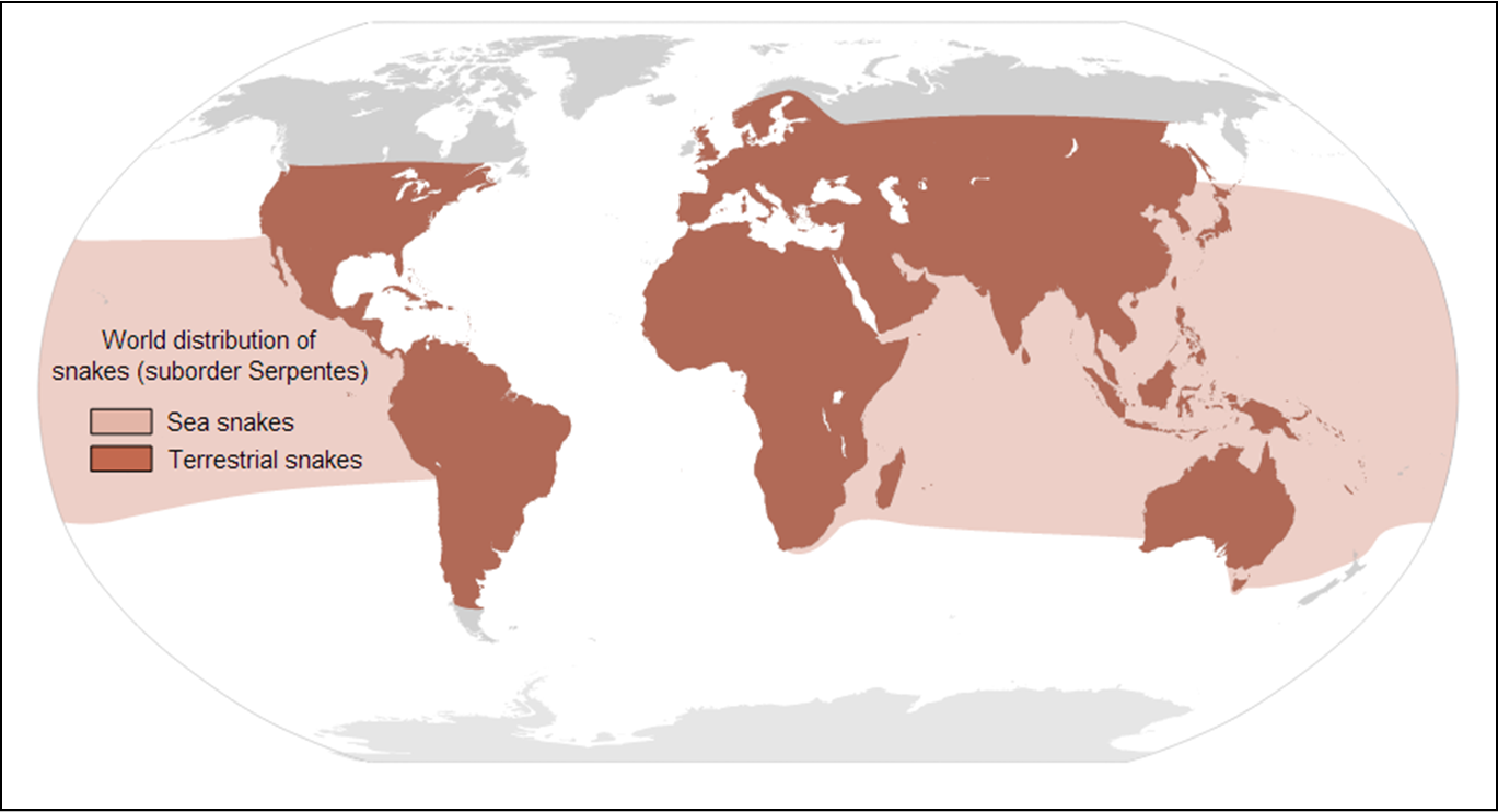
Global distribution of 35 species of the genus Crotalus
Data from IUCN; click for a larger version
Rattlesnakes have captured the attention of Europeans ever since they first started settling the New World. Using their Nahuatl (Aztec) name, Teuhtlacocauhqui, Francisco Hernández described them in his 1615 Quatro Libros de la Naturaleza"When they strike, the bite is fatal unless treated promptly...It has a tail with rattles, one for each year of its life...It has two curved fangs in its upper jaw to inject its venom...It moves in a slithering fashion. Indians hunt and capture them and hang them around their necks...Those who raise them at home say they can live for up to a year without eating anything...When wounded and angry, it whips around, shaking its rattles, and raises its neck to frighten those nearby. However, it does not bite unless provoked." Hernández's book also contains the earliest illustration of a rattlesnake, which is certainly Crotalus durissus. Many other 16th and 17th century authors also wrote about rattlesnakes, dating back to their earliest mention in print, by Cieça de Leon in 1554. Many of these writings contain both accurate information and the first printed iterations of several still-current myths. Clearly, native Americans had known of rattlesnakes since ancient times; the Aztecs and Mayans had a rattlesnake constellation which may have been part of their zodiac.

Top: Neotropical Rattlesnake (Crotalus durissus)
Bottom: The earliest illustration of a rattlesnake
in a book, from Hernández 1628
Because of their tails, Linnaeus thought that rattlesnakes were so unusual that he placed them in their own genus, Crotalus, separating them from other vipers (which he placed in the genus Coluber despite their solenoglyphous fangs). Linnaeus described three species of rattlesnakes: Crotalus horridus (see below), C. durissus, and C. dryinas. Like Boa constrictor and B. orophias, we now consider the latter two species to be the same, but unlike the boa names we use the name durissus for the species even though dryinas precedes it on the page5. Although most rattlesnakes are North American, Linnaeus's C. durissus specimen was collected by Claes Grill in Surinam and originally described in detail in 1748 in the same dissertation that contained the boa constrictor description. In contrast, the "C. dryinas" specimen was from the king of Sweden's collection and lacked geographic data. It is clear that King Adolf Frederick was not the most attentive curator—his curatorial record is incomplete, and over time many of his specimens have been lost and their labels mixed up or deliberately modified. He wasn't the most assertive head of state either, described as "little more than a state decoration"—although apparently he did like snakes. Both rattlesnake specimens from his collection that Linnaeus examined have apparently been lost for over 100 years, and the Grill specimen is lost as well. As a result, and because Linnaeus's descriptions are so terse, the names of the three rattlesnakes (horridus, durissus, and dryinas) were for many years confusingly and inconsistently applied. For example, both Holbrook and Duméril referred to the timber rattlesnake as C. durissus in their respective classic works, Duméril additionally called the neotropical rattlesnake C. horridus, and Boulenger refers to the eastern diamondback as C. durissus in his catalogue of snakes in the British Museum.

Crotalus horridus

Crotalus horridus is the type species of the genus Crotalus,
which today contains 39 species.
Of this snake which would come to symbolize America, Linnaeus wrote (appropriately) "Lives in America. Very venomous; its antidote is Senega (snakeroot). It is eaten by pigs, and calls down birds and squirrels from the trees into its jaws." This is a lengthy description for him, especially considering that timber rattlesnakes had already been described in 1683 (by Edward Tyson, who dissected one), 1721 (by Richard Bradley), 1734 (by Albertus Seba), and in 1745 and 1754 by Linnaeus himself (the first time in a dissertation defended by Barthold Rudolph Hast describing specimens from Count Carl Gyllenborg's collection of rare herps, insects, corals, and minerals, and the second time in his description of the collection of the Swedish king, Museum Adolphi Friderici). C. horridus is the only North American snake Linnaeus described that was not sent to him by Kalm or by his other primary North American informant, Alexander Garden. The specimen that he described had 7 rattle segments, and he was able to give a count of the ventral scales (167), which indicates that he examined a complete specimen, but the specimen that is now in the Royal Museum in Stockholm is represented only by a severed tail, which has 12 rattle segments, and a head, which is actually from a bushmaster (Lachesis muta). Like the specimens of the neotropical rattlesnakes, apparently the original specimen has been missing since at least 1899, and possibly much earlier.6

Catesby's Timber Rattlesnake (which he called Vipera
caudisona
, but which Linnaeus and we call Crotalus horridus).
Kalm considered this "an incomparable illustration".
Although Kalm did not collect a rattlesnake for Linnaeus, he gave a lengthy, detailed, and incredibly accurate description of rattlesnakes and their relationship with humans, which is as much an account of snake biology as it is of the cultural history of colonial America. His words suggest that rattlesnakes were already on their way out in eastern North America in the mid-1700s: "In all my travels, I saw only 3 living specimens. I frequently heard them in the nearby thicket, but it seemed inadvisable to pursue them." It is a testament to Kalm's scientific training that he surpassed many modern observers in accurately stating that "The snake is usually 3 to 4 feet long. The largest one I saw was 6 feet long and as thick as the calf of a man's leg. Usually they are as thick as the wrist" and "They travel slowly, thus one need not fear being overtaken" and "The rattler is peculiar in that he usually does not injure a person unless forced to defend himself."  Despite these honest observations, Kalm had no special love of snakes, including them in a list of reasons that he preferred Sweden to America: "The rattlesnakes, horned-snakes, red-bellied, green, and other poisonous snakes, against whose bite there is frequently no remedy, are in great plenty here".

Timber Rattlesnake from Bradley (1721)
Catesby also described the Timber Rattlesnake, which he called Vipera caudisona, at length. Gmelin, writing in the 13th edition of Systema Naturae, expanded Linnaeus's description in both inaccurate ("The most venomous of the serpent tribe") and accurate ("They seldom bite unless when irritated, or for the purpose of securing their prey, and the fascinating power which has been attributed to them is probably nothing more than that they first bite the animal and patiently watch till it dies to devour it") ways. In 1754 in Museum Adolfi Frederici Linnaeus wrote:"...nor can he escape with life who is bitten by the Rattle-snake (Crotalus horridus) in any part near a great vein. But the merciful God has distinguished these pests by peculiar signs, and has created them most inveterate enemies; for as he has appointed cats to destroy mice, so has he provided the Ichneumon [mongoose] (Viverra Ichneumon) against the [cobra], and the Hog to persecute the latter. He has moreover given the Crotalus a very slow motion, and has annexed a kind of rattle to its tail, by the motion of which it gives notice of its approach; but, lest this slowness should be too great a disadvantage to the animal itself, he has favoured it with a certain power of fascinating squirrels from high trees, and birds from the air into its throat, in the same manner as flies are precipitated into the jaws of the lazy toad."

Other notable snake species described by Linnaeus

Linnaeus described 83 other snake species between his 10th and 12th editions that are still considered valid today, plus 31 that are not (including Crotalus dryinas and Boa orophias). These include many familiar, widespread, and notable species, including 2 scolecophidians, Anilius scytale, and an Asian pipesnake from Sri Lanka (all of which he placed in the genus Anguis, which we today use for legless lizards), several huge constrictors including the Indian Python, Boa Constrictor, and Green Anaconda (but also three smaller tree boas and two sand boas, the latter also in Angius), 13 vipers including the fer-de-lance, copperhead, European adder, bushmaster, and pygmy rattlesnake, a pair of homalopsids, 46 colubrids (including many familiar European and American species but also an African egg-eater and an Asian flying snake), 5 lamprophiids, and 9 elapids (including 3 cobras, 2 coralsnakes, and 2 sea snakes). He also made a few brief comments about snake anatomy and biology as footnotes or in his introductory material, including his method for counting ventral and subcaudal scales (first used in Amphibia Gyllenborgiana and still in use today) as well as the correct observations that "Serpents of our country hibernate and in the early spring shed their skin, that is to say, their old age" and "Serpentes often swallow down prey twice as thick as their neck, on account of their expandable, unarticulated jaws". In other works, he presents a great deal of information on snakebite and, the consummate botanist, its treatment using various medicinal plants. Although Linnaeus bore no special love for snakes, he treated them as he did other biodiversity, and I encourage all modern biologists to do the same—to view snakes as wildlife rather than pests, as a beautiful and diverse part of our natural heritage, to see them as what they are rather than what we imagine them to be.

It is tempting to imagine Linnaeus as a brilliant solitary taxonomist, aided and sent specimens by his correspondents, colleagues, and students but intellectually working alone. But, as today, Linnaeus relied heavily on his network both to obtain specimens and to describe them with reference to those who had gone before. Of the 74 species in the 10th edition, only four were brand new original descriptions (these were Vipera aspisfrom southern Europe, Epicrates cenchria from South America, Erythrolamprus triscalis from Curaçao, and Duberria lutrix from Africa), and the 12th contained scarcely more, mostly southeastern North American species sent to Linnaeus by Alexander Garden. Almost ten times that many new snakes were described last year alone.

Coronella austriaca from Laurenti 1768
It's probably safe to assume that Linnaeus described every snake he ever saw, since this is what he did with everything else. And, considering he lived in Sweden most of his life and never traveled further south than Germany, he did pretty well, nailing numerous tropical species of snake collected by others and sent either to him, or that he examined in the collections of zoologists in Germany, England, and Holland. Systema Naturae contains snakes from every continent except for Australia, which was only just becoming known in Europe at the time of Linnaeus's death (his correspondant Joseph Banks and two of his apostles, Daniel Solander and Anders Sparrman, sailed around the world with James Cook and visited Australia and Oceania in the 1760s and 1770s; Linnaeus's health was poor throughout the 1770s and he died in 1778). But, there is one glaring oversight in Linnaeus's snake work: he described only two of the three native Swedish snakes (Natrix natrix and Vipera berus). Both of these he initially described in his 1746 Fauna Svecica, an account of the animals of Sweden containing 1,357 species in its original edition (upated 1761 with 2,266 species), in which he used cumbersome pre-binomial names such as Coluber natrix scutis abdominalibus CLXX squamis caudae LX ("Water snake with 170 ventral scales and 60 subcaudal scales"), which later became the much simpler yet no less unequivocal Coluber natrix in Systema Naturae. But he missed one: the smooth snake, Coronella austriaca, which was described by J.N. Laurenti7 and named for his native Austria (where it is also found) ten years after the 10th edition of Systema Naturae. Did Linnaeus ever see a Coronella in all the years he lived, worked, and botanized in Sweden? Smooth snakes are active during the day in dry, sunny clearings where they bask in bushes, and although they are not found as far north as Uppsala, they do occur in Småland, where Linnaeus grew up. It seems likely that Linnaeus would have seen them—did he think they were the same species as another kind of snake? If not, why did he leave them out of Fauna Svecica and Systema Naturae, which were intended to be as comprehensive as possible?


1 In the 10th edition Linnaeus confused specimens of racers with those of the black form of the Eastern Hognose Snake (Heterodon platirhinos), but by the 12th edition these had been separated and the phrase "triangular head" removed from the description of the racer.


2 After whom the mountain laurel genus Kalmia is named.


3 Like Maria Sibylla Merian before him, Catesby was among the first naturalists to draw his plants and animals interacting in their natural habitats, a style of representation that would later be used by Alexander Wilson and John James Audubon. He was also the first to abandon the Native American names for his subjects, instead establishing scientific binomials based on relationships a la Linnaeus. Had his work been published three decades later, he might have been immortalized as the father of North American herp taxonomy, and many of the scientific names that we use today could have been very different. Catesby's book, richly illustrated, was much more popular than Linnaeus's.


4 The specimen named Coluber leberis was likely a Storeria, the only genus found in the area traversed by Kalm (Pennsylvania, New York, New Jersey, and southern Ontario) with matching scale counts. Although the scale counts and pattern description match S. occipitomaculata better and this species is more common than S. dekayi in northeastern North America, the specimen could have been either, and since we cannot examine it, the name is not used. Coluber ovivorus is even more enigmatic, because the description does not match any northeastern snake well.


5 This is because, by the time it was all sorted out, the name C. durissus had ended up being in more widespread use, so the "proper" name dryinas was suppressed by the International Commission on Zoological Nomenclature.


6 There is a reasonable chance that the specimen that Linnaeus first named C. horridus was actually from South America, and thus was really C. durissus as well, but since we cannot prove this beyond a shadow of a doubt, in 1926 the International Commission on Zoological Nomenclature decided to continue to use it for the timber rattlesnake.


7 Little is known about Laurenti. No picture of him exists, and his 1768 thesis, 
Specimen medicum, was his only publication. In it, he elevated Linnaeus's order Reptilia to a class, distinguishing it from class Amphibia, into which Linnaeus lumped both amphibians and reptiles. Laurenti also tripled the number of reptile genera, coining some of today's most familiar genus names, including Vipera, Natrix, Laticauda, Dipsas, and Naja.


ACKNOWLEDGMENTS

Thanks to Todd PiersonPatrick Jean, and JD Willson for the use of their photos, and to my mom for getting me William Blunt's Linnaeus for Christmas this year, which inspired this article.

REFERENCES

Andersson, L.G. 1899. Catalogue of the Linnaean type-specimens of snakes in The Royal Museum in Stockholm. Bihang till Kongl. Svenska Vetenskaps-Akademiens Handlingar 24:1-35 <link>

Blunt, W. 2002. Linnaeus: The Compleat Naturalist. Princeton University Press, Princeton, New Jersey, USA <link>

Bradley, R. 1721. A philosophical account of the works of nature, London <link>

Campbell, J.A. and W.W. Lamar. 2004. The Venomous Reptiles of the Western Hemisphere (2 Vol.). Cornell University Press, Ithaca, New York <link>

Catesby, M. 1731. The Natural History of Carolina, Florida, and the Bahama Islands, London.<link>

Cieça de Leon, P. 1554. La Crónica del Perú, Seville <link>

Gronovius, L.T. 1756. Museum Ichthyologicum. Theodorum Haak, Lugduni-Batavorum <link>

Hernández, F. 1615. Quatro libros. De la naturaleza, y virtudes de las plantas, y animales que estan receuidos en el vso de medicina en la Nueua España, y la methodo, y correccion, y preparacion, que para administrallas se requiere con lo que el doctor Francisco Hernandez escriuio en lengua latina <link>

Kalm, P. 1771. Travels Into North America. T. Lowndes, London <link>

Kitchell, K. and H.A. Dundee. 1994. A trilogy on the herpetology of Linnaeus's Systema Naturae X. Smithsonian Herpetological Information Service 100 <link>

Klauber, L.M. 1941. The rattlesnakes listed by Linnaeus in 1758. Bulletin of the Zoological Survey of San Diego 17:81-95 <link>


Klauber, L.M. 1948. Some Misapplications of the Linnaean Names Applied to American Snakes. Copeia 1948:1-14 <link>

Klauber, L.M. 1956. Rattlesnakes: their habits, life histories, and influence on mankind. University of California Press <vol 1/vol 2>

Krecsák, L. and R. Wahlgren. 2008. A survey of the Linnaean type material of Coluber berus, Coluber chersea and Coluber prester (Serpentes, Viperidae). Journal of Natural History 42:2343-2377 <link>

Larsen, E.L. 1957. Pehr Kalm's Account of the North American Rattlesnake and the Medicines Used in the Treatment of its Sting. American Midland Naturalist 57:502-511 <link>

Laurenti, J.N. 1768. Specimen medicum: exhibens synopsin reptilium emendatam cum experimentis circa venena et antidota reptilium austriacorum. Typ. Joan. Thomae nob. de Trattnern, Vienna, Austria <link>

Linnaeus, C. 1745. Amphibia Gyllenborgiana. Uppsala University, Uppsala <link>

Linnaeus C. 1746. Fauna Svecica Sistens Animalia Sveciæ Regni: Quadrupedia, Aves, Amphibia, Pisces, Insecta, Vermes, Distributa per Classes & Ordines, Genera & Species. Differentiis Specierum, Synonymis Autorum, Nominibus Incolarum, Locis Habitationum, Descriptionibus Insectorum. Stockholmiæ [Stockholm] (Sweden): Sumtu & literis Laurentii Salvii <link>

Linnaeus, C. 1746. Museum Adolpho Feidericianum. Uppsala University, Uppsala <link>

Linnaeus, C. 1748. Surinamensia Grilliana. Uppsala University, Uppsala <link>

Linnaeus, C. 1764. Museum S:ae R:ae M:tis Adolphi Friderici Regis Svecorum, Gothorum, Vandalorumque &c. &c. &c. in quo Animalia rariora imprimis & exotica: Aves, Amphibia, Piscis describuntur. Tomi secundi Prodromus. Holmiae. Pp 110. <link/translated>


Linnaeus, C. 1758. Systema naturae per regna tria naturae, secundum classes, ordines, genera, species, cum characteribus, differentiis, synonymis, locis. Tomus I. Editio decima, reformata. Holmiae, 823 pp.<link>


Linnaeus, C. 1762. Morsura Serpentum. Uppsala <link>

Linnaeus, C. 1766. Systema naturae per regna tria naturae, secundum classes, ordines, genera, species, cum characteribus, differentiis, synonymis, locis. Tomus I. Editio duodecima, reformata. Holmiae, 532 pp <link>

Lönnberg, E. 1896. Linnean type-specimens of birds, reptiles, batrachians and fishes. Bihang till Kongl. Svenska Vetenskaps-Akademiens Handlingar 22:1-45 <link>

McIvor, R.S. 2010. Aztec Constellations Preserved by Duran. Journal of the Royal Society of Canada 104:46-50 <link>

Seba, A. 1734-1765. Locupletissimi rerum naturalium thesauri accurata descriptio, et iconibus artificiosissimis expressio, per universam physices historiam :opus, cui, in hoc rerum genere, nullum par exstitit. Apud Janssonio-Waesbergios & J. Wetstenium & Gul. Smith, Amstelaedami <link>

Smit, P. 1979. The zoological dissertations of Linnaeus. Svenska Linnesallskapets Arsskrift 1978:118-136 <link>

Tyson, E. 1683. Vipera Caudi-Sona Americana, Or the Anatomy of a Rattle-Snake, Dissected at the Repository of the Royal Society in January 1682/3 by Edw. Tyson MD Coll. Med. Lond. Cand. & RS Soc. Philosophical Transactions (1683-1775) 13:25-46 <link>

Creative Commons License

Life is Short, but Snakes are Long by Andrew M. Durso is licensed under a Creative Commons Attribution-NonCommercial-NoDerivs 3.0 Unported License.

Snakes that decapitate their food

$
0
0
This article will soon become available in both Spanish!
Este artículo se convertirá pronto disponible en español!

Click here to read this article in Japanese
日本語版の記事はこちら

Crab-eating Snake (Fordonia leucobalia) eating a crab
A few years ago I wrote an article about southeast Asian crab-eating snakes, the only snakes (at the time) known to break apart their food instead of swallowing it whole. Although I ended that article by wondering how many more strange snake dietary adaptations we might discover, I didn't actually anticipate writing a sequel to that article—it was so unique that the BBC filmed it for their series Life in Cold Blood, and I doubted that anyone would discover another snake that tore apart its prey. You can imagine my surprise when recently I was asked to review a paper about another snake that breaks its food apart! I was also delighted that this snake was a scolecophidian, because I feel that they are underrepresented both on this blog and in snake biology in general. It is a bit unsatisfying that it is the Brahminy Blindsnake (Indotyphlops braminus, formerly known as Ramphotyphlops braminus), the best studied scolecophidian by far by virtue of its enormous range and unusual breeding habits, but I think this exciting discovery could become extended to some or most of the other >400 species of blindsnakes.

A blindsnake with decapitated termite heads
stuck to the back of its head
Late last year, herpetologist Yosuke Kojima, a postdoctoral researcher at Kyoto University, and entomologist Takafumi Mizuno, a graduate student at Kyoto Institute of Technology, made a chance finding. They had been close friends since elementary school and shared an interest in behavioral and chemical ecology. Together, they planned some experiments to learn more about interactions between blindsnakes and their primary prey, ants. Mizuno's lab also kept colonies of termites (in this case, Reticulitermes speratus), which are also eaten by blindsnakes. Blindsnakes are unusual in that they eat many small prey at a time rather than a few large prey infrequently. Blindsnakes often eat 20 or more prey items at a time, and the maximum number of prey items ingested by a single individual is 1,431 for Anilios (Ramphotyphlops) nigrescens from Australia. Because blindsnakes often gorge themselves when feeding in an ant or termite nest, they often eat very quickly, using a raking technique of the mandibles (in leptotyphlopids) or of the maxillae (in typhlopids). Nate Kley's lab at Stony Brook University has taken some fantastic videos of blindsnake feeding techniques.

Time-sequence of a blindsnake ingesting and decapitating
a termite worker. From Mizuno & Kojima 2015
Supplementary video here
As Mizuno fed termites to the blindsnakes, he observed something very unusual. The blindsnakes typically grabbed and swallowed the termites backwards. Most snakes usually swallow their prey head-first, so this was weird enough. But, it gets weirder. Often, when the snake had maneuvered a termite so that only its head stuck out of the snake's mouth, it would rub its face on the bottom of the tank, decapitating the termite. All of the termite soldiers and about half of the termite workers offered to the blindsnakes were decapitated. Occasionally, a snake would regurgitate a termite that it had consumed whole, decapitate it, and re-consume the body. Decapitated termite heads became attached to the back of the snake’s head or were scattered around the bottom of the cage. The snakes never ate the decapitated heads. There did not appear to be a cost to decapitation—whether a snake decapitated a termite or not, the time required to completely ingest it was about 3 seconds. However, twice blindsnakes were observed swallowing termites head-first, which took only about 1-1.5 seconds. This may not seem like a big difference, but when you're eating hundreds or thousands of prey items in one sitting, it can add up!

Intact termite heads in the feces of a blindsnake
From Mizuno & Kojima 2015
Why do blindsnakes remove the heads of their prey? One reason might be that termite heads contain glands full of toxic chemicals called terpenes. But, unlike predators that remove the skin of various amphibians to avoid the toxins in their skin glands, blindsnakes don't always remove the heads of their prey, suggesting that they aren't that susceptible to terpene poisoning. It's even been suggested that some blindsnakes might be sequestering defensive chemicals from the ants and termites that they eat, just as gartersnakes sequester tetrodotoxin from newts, in which case they might actually prefer the part of the termite with more chemicals. A more likely hypothesis is that the heads are less digestible than the termites' bodies. Between 26 and 100% of the termite heads consumed by blindsnakes in Mizuno & Kojima's experiment remained undigested in the snakes' feces. Additionally, the snakes preferred to eat the worker termites rather than the more heavily-armored soldier termites, and the few soldier termites they did eat were newly-molted. Removing the termites' scleritized heads might allow blindsnakes to pack more soft, squishy bodies into their stomachs, maximizing the nutrition they get out of their meals. It's a bit like you or me peeling a banana or an orange, or removing the husk from a coconut. Since soldier termites have pinching mandibles, removing their heads might also prevent the blindsnakes from being bitten from the inside, which is a bit like you or me...removing the horns of a cow before eating it, if we ate cows alive and whole, I guess?

Evidently the raking maxillae of typhlopids
are sufficiently dexterous to manipulate
prey inside the mouth to position them
for decapitation.
From Kley 2001
Since snakes don't have hands, they've got to remove any indigestible parts using the only maneuverable part they do have—their jaws. Unlike other blindsnakes (which use bilaterally synchronous jaw movements similar to those of all other vertebrates) but like alethinophidians, typhlopid blindsnakes can move the left and right sides of their highly mobile upper jaws independently and asynchronously. Despite its sophistication, the ratcheting movements of their maxillary raking mechanism are insufficient, by themselves, to allow them to decapitate their prey. We must await further functional-morphological studies to assess the role of the toothless lower jaw, which could act as a wedge or blade, in this process. Since snakes cannot really "bite", arthropods, with their jointed limbs and bodies, might be the only type of prey that a snake could pull apart. There are a fair number of snakes that eat arthropods, but most of them are relatively obscure. Besides the crab-eating snakes, one might look for prey-dismembering behavior in sonorines, a tribe of desert-dwelling snakes from southwestern North America, other North American snakes such as the colubrines Tantilla and Opheodrys and the natricine Regina, the dwarf racers of Africa and the Middle East (genus Eirenis), the centipede-snakes of Africa (genus Aparallactus), or certain kukrisnakes (genus Oligodon). In addition to the typhlopid blindsnake in this study, two short notes from the 1950s and 60s document similar decapitation behaviors in two different species of leptotyphlopids (Epictia goudotii [formerly Leptotyphlops phenops] from Central America and Rena dulcis [formerly L. dulcis] from Texas), despite their radically different jaw morphology. I won't be surprised if it turns up in other scolecophidian families as well, since this most-basal group of living snakes probably co-evolved with the early radiation of ants and termites, their favorite prey.

ACKNOWLEDGMENTS

Thanks to Brendan Schembri for the use of his photo, and to Takafumi Mizuno and Yosuke Kojima for giving me the opportunity to write about their discovery in advance of its publication and for translating it into Japanese.

REFERENCES

Kley, N.J. 2001. Prey transport mechanisms in blindsnakes and the evolution of unilateral feeding systems in snakes. American Zoologist 41:1321-1337 <link>

Mizuno, T. and Y. Kojima. In press. A blindsnake that decapitates its termite prey. Journal of Zoology 10.1111/jzo.12268 <link>

Prestwich, G.D., B. Bierl, E. Devilbiss, and M. Chaudhury. 1977. Soldier frontal glands of the termite Macrotermes subhyalinus: Morphology, chemical composition, and use in defense. Journal of Chemical Ecology 3:579-590 <link>

Reid, J.R. and T.E. Lott. 1963. Feeding of Leptotyphlops dulcis dulcis (Baird and Girard). Herpetologica 19:141-142  <link>

Savitzky, A.H., A. Mori, D.A. Hutchinson, R.A. Saporito, G.M. Burghardt, H.B. Lillywhite, and J. Meinwald. 2012. Sequestered defensive toxins in tetrapod vertebrates: principles, patterns, and prospects for future studies. Chemoecology 22:141-158 <link>

Shine, R. and J.K. Webb. 1990. Natural history of Australian typhlopid snakes. Journal of Herpetology 24:357-363 <link>

Smith, H.M. 1957. Curious feeding habit of a blind snake, Leptotyphlops. Herpetologica 13:102 <link>

Stokes, A.N., A.M. Ray, M.W. Buktenica, B.G. Gall, E. Paulson, D. Paulson, S.S. French, E.D.B. III, and J. E.D. Brodie. 2015. Otter predation on Taricha granulosa and variation in tetrodotoxin levels with elevation. Northwestern Naturalist 96:13-21 <link>

Vidal, N., J. Marin, M. Morini, S. Donnellan, W.R. Branch, R. Thomas, M. Vences, A. Wynn, C. Cruaud, and S.B. Hedges. 2010. Blindsnake evolutionary tree reveals long history on Gondwana. Biology Letters 6:558-561 <link>

Creative Commons License

Life is Short, but Snakes are Long by Andrew M. Durso is licensed under a Creative Commons Attribution-NonCommercial-NoDerivs 3.0 Unported License.

Basics of Snake Fangs

$
0
0

Solenoglyphous fangs of a Gaboon Viper
Snake fangs are specialized, elegantly modified teeth. Some are like hypodermic needles, others are more like water slides. But all serve essentially the same purpose: to inject venom into the snake's prey. Occasionally, the fangs are also used in defense, but studies show that snakes striking in defense are far less likely to inject venom than when they're striking at a prey item, a fact that has assuaged the fears of many an ophidiophobic. I wanted to write a brief review of snake fang types, because their anatomy is very interesting and also because of their important role in classifying snakes and understanding how different groups of snakes are related to each other.

Cross-sections of fangs:
F is an aglyphous tooth.
G is an opisthoglyphous fang.
H is a proteroglyphous fang.
I is a hollow solenoglyphous fang.
From Bauchot (2006)
Many snakes produce venom, which is essentially very strong saliva, in glands in their heads (which is where you produce your saliva, too). We call these glands venom glands if they are well-developed, complete with an interior cavity, a duct connecting to a hollow fang, and compressor muscles that generate high pressures when the jaws are rapidly closed. If they lack these features, we usually call them Duvernoy's glands instead. Because there is a lot of variation among snake species in the structure of these glands and their associated teeth, there is some debate about whether or not venom glands and Duvernoy's glands are really two forms of the same thing. Either way, three groups of snakes (atractaspidines, elapids, and viperids) have independently evolved an advanced apparatus to deliver large quantities of venom during a brief strike, and many other snakes (and a few lizards) have evolved less sophisticated, but still relatively effective, modifications to their teeth in order to deliver venom after they have grabbed their prey and are "chewing" on it. The teeth of modern snakes are classically divided into four types, three of which are typically called fangs. The four tooth types have fancy names, all of which involve the Greek word glyph, one of the meanings of which is "groove". They are as follows:

Solenoglyphous

Folding of solenoglyphous fangs.
Fang is in red, maxilla green,
prefrontal orange, pterygoid yellow,
ectopterygoid purple. Vipers lack
premaxillary and palatine teeth.
From Bauchot (2006)
This most sophisticated fang type evolved once, in the ancestor to all modern vipers, which lived in Asia about 40 million years ago. Fossils suggest that solenoglyphous fangs have changed little since that time, even though vipers have undergone an incredibly successful radiation into 320 extant species found on all continents except for Australia and Antarctica. Solenoglyphous fangs are long and tubular and are attached to the snake's maxillary bone. Most snakes have several tooth-bearing bones, including four (the premaxilla, maxilla, pterygoid, and palatine) in the upper jaw, and one (the dentary) in the lower. In humans, three of these bones (the premaxilla, maxilla, and dentary) also bear teeth - your premaxilla holds your top incisors, while your maxilla holds your upper canines and molars and your dentary all your lower teeth - while the others form part of the roof of the mouth. In vipers, the maxilla bears only a single tooth (the fang) and is hinged so that the fangs can be folded back parallel to the jaws when the mouth is closed, or erected perpendicular to the jaws, the position when striking. The teeth in the pterygoids and dentaries work together to manipulate food once it gets into the mouth. Solenoglyphous fangs are strikingly similar to hypodermic needles. They have a hollow core that receives venom from the venom gland at the entrance orifice near the base and injects it from a slit-like exit orifice on the front of the fang near the tip. If the opening were at the very tip of the fang, its strength would be compromised and it would lack the sharp point needed to penetrate the target. Even under normal use, vipers shed their fangs every two months.
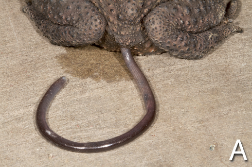
Modified solenoglyphous fang of
African Burrowing Asp (Atractaspis engaddensis)
A similar fang type evolved a second time about 29 million years ago in a group of African snakes, currently placed in the family Lamprophiidae, subfamily Atractaspidinae. Two genera, Atractaspis(mole vipers, burrowing asps, or stiletto snakes) and Homoroselaps (African dwarf garter or harlequin snakes), possess elongate anterior fangs, although only those of the stiletto snakes are movable. Stiletto snake fangs pivot on a socket-like joint that is more flexible than those of vipers, allowing these snakes to strike beside and behind them with their mouth closed. This is an adaptation to living underground and envenomating small mammals and other reptiles in narrow subterranean burrows. The fang morphology of atractaspidines and viperids is remarkably similar, considering that these two snake lineages last shared a common ancestor over 40 million years ago.

Proteroglyphous

Proteroglyphous fangs of an Eastern Green Mamba
(Dendroaspis angusticeps). Don't try this.
From Bauchot (2006)
This fang type also evolved only once, in the ancestor to all modern elapids, which lived 25-40 mya in Asia or Africa. Proteroglyphous fangs are in the front of the mouth and are about three times shorter than solenoglyphous fangs. This is because they are not hinged. Snakes with proteroglyphous fangs typically strike their prey and hang on until the venom has taken effect, as opposed to releasing they prey and then tracking it down. Some elapids constrict their prey at the same time as envenomating it. Over 350 species of elapids exist today, including well-known groups such as cobras, mambas, death adders, taipans, coralsnakes, and sea snakes, and less-well-known species, mainly found in Australia, of which a good number are small, secretive, and not considered dangerous to humans.

Maxilla of a proteroglyphous snake showing the almost
completely closed groove along the anterior edge connecting
the two orifices, as well as the aglyphous tooth at the
rear of the maxilla. This line may be obscured in longer fangs.
From Shea et al. 1993
Unlike solenoglyphs, some proteroglyphs have other teeth on the maxilla behind the fang. However, the fang is always separated from the other teeth by a gap, called a diastema. Some elapids have more than one functional fang on each side. In both vipers and elapids, there are usually at least two fangs on each maxilla at any one time, one that is in use and one that is a reserve fang. Both fangs are draped in a layer of connective tissue and skin called the fang sheath. Some proteroglyphs have partially movable fangs, including many of the most dangerous species such as mambas, taipans, and death adders. A few, such as spitting cobras, have modified exit orifices to their fangs that are smaller and rounder than in other cobras, a modification that increases the velocity with which venom is ejected. Modifications to the muscles and the fang sheath also facilitate spitting in these cobras. A few elapids, such as sea snakes that eat only fish eggs, have lost their fangs and their venom glands, which suggests that the primary role of venom, at least among elapids, is in feeding rather than in defense.

Opisthoglyphous

Opisthoglyphous fang of Eastern Hog-nosed Snake
These are commonly known as "rear-fanged" snakes. Opisthoglyphous fangs are grooved rather than hollow and are found near the back of the maxilla, behind the normal teeth. Typically, snakes with rear fangs must chew on their prey to bring their fangs into a biting position. There is considerable variation in the size, shape, and number of opisthoglyphous fangs from species to species, and sometimes even within a species. Most opisthoglyphous fangs are connected to Duvernoy's glands, which differ from true venom glands in several important ways, most notably in that they lack associated muscles to generate the pressure needed to evacuate venom, as in solenoglyphous and proteroglyphous snakes. The pressure on the venom glands of biting solenoglyphs and proteroglyphs can exceed 30 psi, the pressure of a car tire, whereas the pressure inside the Duvernoy's glands of opisthoglyphs is generally less than 5 psi. Because Duvernoy's glands also lack a chamber for storing venom, the idea is emerging that opisthoglyphous snakes probably secrete their venom only during chewing, which explains why prolonged bites by opisthoglyphs generally have more severe effects.

Opisthoglyphous fangs of Boomslang (Dispholidus typus)
Don't do this either.
Most of these snakes are not harmful to humans, with a few notable exceptions. Boomslangs and Twigsnakes are arboreal, diurnal African colubrines that prey on lizards and birds. They have short heads, rear fangs situated comparatively close to the front of the mouth, and partially muscled Duvernoy's glands. They also have potent venoms and their bites have killed several people, including two prominent snake biologists, Karl Schmidt and Robert Mertens. Bites from other rear-fanged snakes are known to cause relatively mild, transient, and local symptoms, but clinical documentation of these bites and their effects is scattered, incomplete, and frequently anecdotal. Many are written by the victim themselves! The above notwithstanding, bites from opisthoglyphs are generally less medically important than those from proteroglyphs and solenoglyphs. As a result, snake venom research has not focused on them, so there is still much that we do not know about this group of snakes, some of which are becoming increasingly common in the pet trade. Based on what little we do know, the composition of opisthoglyph venom/Duvernoy's secretion is fairly similar to that of viperids, elapids and atractaspidines, which makes sense given that each of these groups is more closely related to certain opisthoglyphs than they are to one another.

A: python, B: viper, C: rear-fanged colubroid, D: cobra
The f  marks the portion of the maxilla where the fang develops.
E shows the elongation of the posterior part of the
maxilla pushing forward the developing fang of a
night adder (d.a.o. = days after oviposition)
From Vonk et al. 2008
Unlike the first two groups, opisthoglyphous fangs appear to have evolved more than once, in snakes as diverse as Quill-snouted Snakes, Neck-banded Snakes, and Boomslangs. At least, that's what we used to think. Actually, it is likely that both solenoglyphous and proteroglyphous fangs evolved from opisthoglyphous fangs, as revealed by an ingenious study that used evidence from embryology and genetics to reveal the evolutionary origins of the three types of snake fangs. In a snake embryo, tubular fangs are formed by the infolding of ridges on the front and back sides of the fang, such as those that form the groove of opisthoglyphous fangs. Furthermore, front fangs develop from the rear part of the upper jaw, and are strikingly similar in their formation to rear fangs. They are pushed into the front of the mouth by disproportionate growth of the initially small part of the maxilla that is behind them. Finally, in the anterior part of the maxilla of front-fanged snakes, expression of a gene called sonic hedgehog, which is responsible among other things for the formation of teeth, is suppressed.


Relative size of the venom gland (VG) in
A: rear-fanged colubrid (Helicops leopardinus),
B: boomslang, C: homalopsid,
D: cornsnake, E: African egg-eater
SG = supralabial salivary gland
From Fry et al. 2008
Although developmental similarity is not conclusive proof of structural homology (similarity due to inheritance rather than due to other factors), these findings are consistent with the hypothesis that solenoglyphous, proteroglyphous, and at least some opisthoglyphous fangs are homologous structures. The hypothetical evolutionary trajectory was thus: some snakes evolved grooved fangs in the rear of their mouth. In a few cases (viperids, elapids, and atractaspidines), they subsequently lost the preceding teeth as what was formerly a rear fang became a tubular front fang. Other snakes retained their anterior teeth (at least some non-front-fanged colubroids), and still others developed fangs but then lost them (aglyphs such as ratsnakes). Evidence for this surprising final part comes from the formation of the maxilla and its teeth, which takes place in a single piece in pythons, but from two pieces in all fanged snakes as well as in ratsnakes, a pattern which supports a single evolutionary origin and subsequent loss of fangs. Additionally, vestigial Duvernoy's glands have been found in ratsnakes, egg-eaters, pareatid slug-eaters, and other nonvenomous aglyphs, a discovery that has led to the misleading generalization that all snakes are venomous and much subsequent misunderstanding among the non-scientific community. Toxic saliva does not a venomous animal make, as evidenced by the fact that even human saliva injected subcutaneously will produce pain and swelling.

Aglyphous

Both boas and pythons have only
aglyphous teeth, which is about
the only thing this film got right.
This word is used to describe unmodified teeth, essentially non-fangs. All snakes, even those that possess fangs of the first three types, have aglyphous teeth which they use for gripping their prey as they manipulate it during swallowing. As I just mentioned, many advanced snakes that today have only aglyphous teeth probably evolved from fanged ancestors. Several of these snakes, such as North American kingsnakes, ratsnakes, and bullsnakes, have atrophied Duvernoy's glands that lack toxin-producing serous cells. These snakes employ other sophisticated techniques, such as constriction, which is also used by more primitive snakes like boas and pythons (which did not evolve from fanged ancestors).

There are very few dangerous species of aglyphs, but one, Rhabdophis tigrinus, is becoming well-known as one of the only snakes capable of sequestering toxins from its prey for use in its own defense. This species has enlarged posterior maxillary teeth that lack grooves, so they are by definition aglyphous. However, it has relatively potent venom and has caused the deaths of several people. Among colubroids, the distinction between opisthoglyphs and aglyphs has never been entirely clear, but I'm distinguishing between them here because they are two of the four traditionally recognized types of snake teeth. Although the four types of snake teeth in this article are commonly discussed, a more accurate classification for snake teeth might be to divide them into tubular (the fangs of viperids, elapids, and atractaspidines), grooved (the rear fangs of non-front-fanged colubroids), and ungrooved (all other snake teeth).

Aglyphous (ungrooved) teeth and rear fangs of
Rhabdophis tigrinus
From Mittleman & Goris 1974
Happily for snake biologists like myself, the evolution of fangs opened the door for a massive evolutionary radiation of advanced snakes (>2800 species, or >80% of all living snake species). Although sophisticated venom delivery systems, of which fangs are just one of many integral parts, were clearly evolutionary advantageous, they have obviously also been costly at times, leading to their loss in ratsnakes, egg-eaters, and other lineages of advanced snakes. Also worth noting is that many lineages of basal snakes have got along just fine without venom, so there is not an inherent superiority of being venomous as the word "advanced" seems to imply. Rather, some have suggested that during periods of transition from forest to grassland, such as that which took place simultaneous to the dramatic colubroid radiation during the Miocene, snake taxa that were characterized by slow locomotion and constriction (boas & pythons) were supplanted by those characterized by rapid locomotion (many aglyphous colubrids) or passive immobilization (tubular- and grooved-fanged vipers, elapids, and atractaspidines that could use venom to catch their prey). Of course, both slow locomotion and constriction have subsequently been re-evolved among the colubroids, but there has been a lot of time since the Miocene.

ACKNOWLEDGMENTS

Thanks to Daniel Rosenberg (boomslang fang) and Nick Kiriazis (hognose fang) for use of their photographs.

REFERENCES

Bauchot R, editor. 2006. Snakes: A Natural History. New York, New York: Sterling Publishers. <link>


Cundall, D., (2002) Envenomation strategies, head form, and feeding ecology in vipers. In: Biology of the Vipers: 149-162. G. W. Schuett, M. Höggren, M. E. Douglas & H. W. Greene (Eds.). Eagle Mountain Publishers, Eagle Mountain, UT <link>


Greene, H. W. (1997) Snakes: The Evolution of Mystery in Nature. Berkeley: University of California Press <link>


Fry BG, Scheib H, van der Weerd L, Young B, McNaughtan J, Ramjan SR, Vidal N, Poelmann RE, Norman JA, 2008. Evolution of an arsenal: structural and functional diversification of the venom system in the advanced snakes (Caenophidia). Mol Cell Proteomics 7:215-246 <link>


Hayes, W. K., S. S. Herbert, G. C. Rehling & J. F. Gennaro, (2002) Factors that influence venom expenditure in viperids and other snake species during predator and defensive contexts. In: Biology of the Vipers: 207-234. G. W. Schuett, M. Höggren, M. E. Douglas & H. W. Greene (Eds.). Eagle Mountain Publishers, Eagle Mountain, UT <link>


Jackson K, 2002. How tubular venom‐conducting fangs are formed. J Morphol 252:291-297 <link>


Kardong, K. V. & T. L. Smith, (2002) Proximate factors involved in rattlesnake predatory behavior: a review. In: Biology of the Vipers: 253-266. G. W. Schuett, M. Höggren, M. E. Douglas & H. W. Greene (Eds.). Eagle Mountain Publishers, Eagle Mountain, UT <link>


Kardong KV, 1996. Snake toxins and venoms: an evolutionary perspective. Herpetologica 52:36-46 <link>


Kuch, U., J. Müller, C. Mödden & D. Mebs (2006). Snake fangs from the Lower Miocene of Germany: evolutionary stability of perfect weapons. Naturwissenschaften 93, 84-87


LaDuc, T. J., (2002) Does a quick offense equal a quick defense? Kinematic comparisons of predatory and defensive strikes in the Western Diamond-backed Rattlesnake (Crotalus atrox). In: Biology of the Vipers: 267-278. G. W. Schuett, M. Höggren, M. E. Douglas & H. W. Greene (Eds.). Eagle Mountain Publishers, Eagle Mountain, UT <link>


Mittleman M, Goris R, 1974. Envenomation from the bite of the Japanese colubrid snake Rhabdophis tigrinus (Boie). Herpetologica 30:113-119 <link>


Pyron, R. A., F. T. Burbrink, G. R. Colli, A. N. M. de Oca, L. J. Vitt, C. A. Kuczynski & J. J. Wiens (2011). The phylogeny of advanced snakes (Colubroidea), with discovery of a new subfamily and comparison of support methods for likelihood trees. Mol. Phylogenet. Evol. 58, 329-342 <link>


Savitzky AH, 1980. The role of venom delivery strategies in snake evolution. Evolution 34:1194-1204 <link>


Shea G, Shine R, Covacevich JC, 1993. Elapidae. In: Glasby C, Ross G, Beesley P, editors. Fauna of Australia. Canberra: AGPS <link>


Vonk FJ, Admiraal JF, Jackson K, Reshef R, de Bakker MA, Vanderschoot K, van den Berge I, van Atten M, Burgerhout E, Beck A, 2008. Evolutionary origin and development of snake fangs. Nature 454:630-633 <link>


Weinstein SA, Warrell DA, White J, Keyler DE, 2011. "Venomous" Bites from Non-Venomous Snakes: A Critical Analysis of Risk and Management of "Colubrid" Snake Bites. Amsterdam: Elsevier <link>


Weinstein SA, White J, Keyler DE, Warrell DA, 2013. Non-front-fanged colubroid snakes: A current evidence-based analysis of medical significance. Toxicon. 69, 103-113 <link>


Weinstein S, White J, Westerström A, Warrell DA, 2013. Anecdote vs. substantiated fact: the problem of unverified reports in the toxinological and herpetological literature describing non-front-fanged colubroid (“colubrid”) snakebites. Herpetological Review 44:23-29.


Wüster, W., L. Peppin, C. Pook & D. Walker (2008). A nesting of vipers: Phylogeny and historical biogeography of the Viperidae (Squamata: Serpentes). Mol. Phylogenet. Evol. 49, 445-459 <link>


Young BA, Dunlap K, Koenig K, Singer M, 2004. The buccal buckle: the functional morphology of venom spitting in cobras. J Exp Biol 207:3483-3494 <link>


Creative Commons License

Life is Short, but Snakes are Long by Andrew M. Durso is licensed under a Creative Commons Attribution-NonCommercial-NoDerivs 3.0 Unported License.

50,000 Hits & Snakes from Florida

$
0
0

Brown Anole (Anolis sagrei)
The purpose of my trip
This week I am in northeast Florida collecting lizards for my PhD research (don't tell anybody who still thinks I only work on snakes). This is a special place for me because it is where I started writing this blog a year and a half ago. Since that time Life is Short but Snakes are Long has received thousands of visitors: almost 100,000 if you go by the stats included with Blogger, but probably closer to 48,000 using stats from the more conservative Google Analytics, which doesn't count bots and other non-human visitors. The true number is probably somewhere in the middle. With many thanks to Alvaro Pemartin and Estefania Carrillo, all posts are available in English and Spanish (the links to the Spanish versions are at the top of each post), and I am working on converting the format of the Spanish pages from PDF to HTML to more closely resemble the English pages. Readers from the USA make up the majority of visitors, but the UK, Canada, Australia, and India are also well-represented, and readers from 177 countries or territories have visited. 

Map of visits to this blog
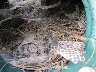
I am proud to have been able to disseminate knowledge about snakes to so many people. The first post on snake sheds is still the most popular, garnering between 44 and 100 hits a day and appearing in the top 10 hits for Google searches for 'snake shed' and 'snake sheds'. Its popularity prompted me to write another article that was less storytelling and more detail about the processes used in snake shed identification. As proof positive that it works, last week I received images of a snake shed from Jean in Lawrence, Kansas, who wrote:

The first photo
I happened across your blog while searching for a way to identify a snake species by it's shedded skin.

We found this [snake shed] in our barn near Lawrence Kansas. I had this extreme fear of snakes so I became proficient in identifying them, if I see them. We have only seen 3 types of venomous snakes in our area, the timber rattler, the western massasauga, and the osage copperhead. Unfortunately, I find that I am truly inept at identifying them by their skins.

We have seen more poisonous snakes this year than usual and we found this skin inside our barn. It very easily could have been trapped inside as we close it up every other evening. We primarily use the barn for storage and workshop. Hopefully, we have allowed plenty of opportunity for the snake to escape.

I mainly want to know if you can help me to identify whether this is a poisonous snake. After reading your blog I am concerned is that it is possibly a copperhead and that it could be hiding. There are numerous places for a creature to stay hidden in our 70 ft barn and I fear that I will open a bin or cabinet and find it, dead or alive.

We love our wildlife and try to be protective and careful, but it seems we have failed at this lately as we recently had to scare an endangered skink out of the barn.

I would appreciate your assistance in possibly identifying this snake. I don't think we have the tail end of the skin. We do have the fairly intact head portion of the skin and can send more pics if needed.

Your blog is very informative and I learned a great deal from it. I thank you in advance for your assistance.

Although the first photo wasn't detailed enough, she was able to find the tail and I was able to help her identify it as a harmless ratsnake, after which she wrote:


The second photo,
showing divided subcaudals
Thank you so much! I checked your blog to take a double look at your pics there and was still unsure, so thank you so much! We did see a few rat snakes earlier this year so my guess is you are spot on!

It is still scary that we didn't see it! We live in a rural area very near to public hunting and fishing but don't have a lot of traffic. It makes my blood boil at the number of snakes we see dead on the SIDES of the roads!

Please keep up the great work! Yours was the first site when I googled snake skin id and by far the most informative i found! I learned so much by reading your blog and I really feel that people need more education about snakes!

Identifying snake sheds has been a new challenge for me. I probably wouldn't have gotten so much practice at it if I hadn't started this blog. I am working on a lot of new content, but I particularly want to develop content that people will find useful and interesting. With that in mind, here are a couple upcoming articles that I've planned:

  • Basics of snakebite
  • Venomous bites from "non-venomous" snakes
  • Common urban snakes
  • Snake predators
  • Invasive snakes
  • Some personal stories about how I became interested in herpetology
  • Several taxon-specific posts
I'm open to suggestions about how to prioritize these and I'm especially open to ideas from readers about new posts that aren't on this list. Some of the best ones I've written so far are ones people have suggested to me. I'm also open to hosting guest posts if there are any interested guest authors out there. Feel free to leave a comment or to contact me by email.

Cornsnake from the island
I also wanted to share a couple of stories from this week. Yesterday we found a young Cornsnake on one of our islands when one of us chased a lizard into the tree hollow where it was hiding. That snake had eaten one of the Brown Anoles in our study, a large male that we marked back in 2011. Young cornsnakes are particularly fond of lizards and ambush them from hiding spots under bark and within decaying trees. My former student David Delaney, now in the Warner lab at UAB, will be conducting research on the effects of cornsnake predation on anole sleeping site selection. I thought this would be the coolest find of the whole trip, but today some folks alerted us to the presence of an Eastern Diamondback Rattlesnake on a public beach right near where we were collecting mainland lizards for David & Dan's lab experiments. This snake was in the surf, which was really foamy due to the wind. The lady who found it said she almost stepped on it. Usually when someone tells you they saw a rattlesnake nearby it's either not a rattlesnake or not there or both, but this time it was for real! I have read about EDBs entering the ocean occasionally, but apparently it is fairly rare. My friend Kerry Nelson, who worked as a naturalist on Little St. Simons Island in Georgia for almost two years and saw diamondbacks in the sand dunes daily, said to me that he never saw one in the surf.

Me with Diamondback
ACKNOWLEDGMENTS

Thanks to Hans Hillewaert, Dan Warner, and Jean Ostrander for their photos and to Jean Ostrander for allowing me to reprint her email.


Creative Commons License

Life is Short, but Snakes are Long by Andrew M. Durso is licensed under a Creative Commons Attribution-NonCommercial-NoDerivs 3.0 Unported License.

How snakes see through closed eyes

$
0
0
Early American symbols depicting rattlesnakes:

(top) Rattlesnake on the $20 bill issued in 1778 by Georgia.
The Latin motto (Nemo me impune lacesset) means,
"No one will provoke me with impunity."

(middle) Benjamin Franklin's "Join or Die" cartoon,
first published in the Pennsylvania Gazette in 1754.
Franklin advocated a rattlesnake as the national symbol
by writing: "...her eye excelled in brightness...she has no
eye-lids. She may therefore be esteemed
an emblem of vigilance."

(bottom) The Gadsden flag, used during
the American Revolution

Normally when someone asks me how to tell the difference between a snake and a legless lizard, I tell them to look at the eyes. Lizards have eyelids whereas snakes do not. Whenever I say this I am lying, although really I am just oversimplifying for the sake of clarity. Most lizards have obvious movable eyelids and so can blink like we do. Snakes, by contrast, seem not to have eyelids. They are ever-staring, ever-vigilant. Ben Franklin esteemed the rattlesnake as a symbol of vigilance because its eyes were always open.

Snakes' eyes are closed all the time. Rather than having movable eyelids, snakes have a single, fused, clear layer of skin over their eye, called a spectacle or brille (German for "glasses"), which protects the eye. A snake's skin is covered in scales, and the outer part of the spectacle is indeed a scale. The deeper layers of the spectacle are formed, during development, from the same embryonic tissue that in other animals forms the eyelid. The spectacle is not attached to the snake's eye in any way, so the eye can move freely behind it, although its movement is limited. This limited movement is because snakes are probably descended from fossorial lizard ancestors that lived underground and had degenerate eyes, much like today's amphisbaenians, although fossil evidence for this hypothesis is scant (as are snake fossils in general).


Eye of an Eastern Ribbonsnake (Thamnophis sauritus)
during the phase prior to shedding when fluid
has built up between the old and new spectacles
Unlike other animals' eyelids, snakes' spectacles are transparent, like a window in their skin, allowing them to see out through their always-closed eyelids. Just before a snake sheds its skin, a layer of fluid builds up between the new inner skin and the old outer layer, clouding the spectacle and causing the other scales to have a faded, milky appearance. This period usually lasts a few days, during which snakes have difficulty seeing and usually will not eat. People who keep snakes as pets have observed that they may become particularly ornery during this period, perhaps as a result of not being able to see clearly.

The horizontal, key-shaped pupil
of Ahaetulla prasina
Another obstacle to snake vision that has been long known but little studied is that snakes' spectacles are vascularized, meaning that they have blood vessels running through them. It is very unusual for tetrapods to have blood vessels in a place that might interrupt their field of vision. First noticed in 1852, these vessels are small but symmetrically distributed across the optically transmissive region of the eye in most species, although the arrangement is radial in basal snakes, acrochordids, and vipers but vertical in colubrids and elapids. In one visually-oriented species, the Asian vine snake (Ahaetulla nasuta), these blood vessels are less dense in the region of the field of vision known as the fovea, where the maximum sharpness is achieved. Most snakes don't have foveas, suggesting that the unusual arrangement of blood vessels in the eyes of Ahaetulla is an adaptation to minimize visual disturbance in this region of highest visual acuity.

Until recently, no one had considered whether movement of blood into and out of the spectacle blood vessels might aid snakes in being able to see. In an article published this week in The Journal of Experimental Biology, Kevin van Doorn and Jacob Sivak of the University of Waterloo in Ontario presented the first evidence that snakes are able to do this. When van Doorn was investigating the mechanisms snakes' eyes use to focus, he noticed the blood vessels in the spectacle, which led him to look more closely at their function. He found that coachwhips, another highly visual species, were able to reduce blood flow to the spectacle in the presence of a potential threat. At rest and undisturbed, newly oxygenated blood flowed into the spectacle blood vessels of the coachwhips for about a minute at a time, interspersed with approximately two minute periods during which no flow took place. When an experimenter walked into the room to perform some routine tasks, spectacle blood flow was almost completely cut off. What little flow there was during this period occurred in short spurts of around 30 seconds each, about half the length of the flow period in undisturbed coachwhips. When the experimenter left the room, the pattern of blood flow in the snakes' eyes returned to normal almost immediately.

Figure from van Doorn & Sivak 2013 showing blood vessels in the spectacle of a Coachwhip (Masticophis flagellum). (A) Image taken during the renewal phase of the integument when the spectacle becomes cloudy. The vessels are most apparent in the region that overlays the iris–pupil boundary because of their higher contrast with the background in this region. (B) The spectacle under retro-illumination, showing the vessels in the illuminated anterior portion of the pupil on the right side. The vessels are dorso-ventrally arranged as is typical for colubrid snakes. Debris and scratches are visible on the spectacle scale (particularly the left side), attesting to its protective role.
Shed skin of a Cornsnake (Pantherophis guttatus)
showing the shed spectacles
Furthermore, van Doorn & Sivak found that when snakes were handled they cut off blood flow to the spectacle completely, probably as part of a sympathetic nervous response. In contrast, blood flow to the eye was continuous and uninterrupted, even during handing, in shedding snakes. You can see a video of blood flow in the spectacle of a shedding corn snake here. Although no experimental evidence has been gathered that filled blood vessels in the spectacle reduce a snake's ability to see, it seems likely given that the blood vessels themselves are quite difficult to see when they are not filled with blood. Snakes actually have remarkably good color vision, better than that of rats and on par with the visual acuity of a cat. Because they move their eyes so little compared to humans, they might be less likely to notice the interruption to their visual field by these blood vessels.

Geckos and some other lizards also have spectacles. A few other species of tetrapods have blood vessels in their optical path, including manatees, armadillos, and some blind salamanders, none of which are renowned for their visual prowess. Little is known about the images these vessels might project onto the vision of these animals, but because they are part of the cornea and so move about with the eye rather than remain stationary relative to it, their area of occlusion would appear to remain stationary to the animal. This is not true for animals with nictitating membranes (diving animals such as penguins or crocodilians) or those with spectacles, both of which have the potential to interrupt the animal's vision. We don't know yet how crocodilians and geckos deal with this issue, but as with so many other features of their lives, snakes have evolved an ingenious and potentially unique solution to a vexing problem, allowing them to remain vigilant as well as keep their eyes protected. Snakes have guarded the Golden Fleece in the Greek tale of the hero Jason and his band of Argonauts, a treasure chamber beneath an ancient city in Rudyard Kipling’s The Jungle Book, and various other treasures in Hindu, Inca, and Basque mythology, all with their eyes closed.

ACKNOWLEDGMENTS

Thanks to Hans Breuer and Kwok Wai for their photographs of Ahaetulla prasina.

Correction: I originally said that the fovea work was done on Ahaetulla prasina, but it was actually Ahaetulla nasuta. Both species have horizontal pupils so it is likely that the reduction in blood vessels is found in both.

REFERENCES

Baker, RA, TJ Gawne, MS Loop, and S Pullman (2007) Visual acuity of the midland banded water snake estimated from evoked telencephalic potentials. J. Comp. Physiol. A 193, 865-870 <link>

Crump, ML (in press) Amphibians, Reptiles, and Humans: Cultural Perceptions and Conservation Consequences. University of Chicago Press.

Foureaux, G, MI Egami, C Jared, MM Antoniazzi, RC Gutierre, and RL Smith. (2010) Rudimentary eyes of squamate fossorial reptiles (Amphisbaenia and Serpentes). Anat. Rec. (Hoboken) 293, 351-357 <link>

Franklin, B (1775) The rattlesnake as a symbol of America. Pennsylvania Journal. <link>

Lüdicke M, 1969. Die kapillarnetze der brille, der iris, des glaskörpers und der chorioidea des auges vom baumschnüffler Ahaetulla nasuta Lacepede 1789 (Serpentes, Colubridae). Zoomorphology 64:373-390.

Mead, AW (1976) Vascularity in the reptilian spectacle. Invest. Opthalmol. Vis. Sci.15, 587-591 <link>

Neher, EM (1935) The origin of the brille in Crotalus confluentus lutosus (Great Basin rattlesnake). Trans. Am. Ophthalmol. Soc. 33, 533-545 <link>

Quekett, J. (1852). Observations on the vascularity of the capsule of the crystalline lens, especially that of certain reptilia. Trans. Microsc. Soc. Lond. 3, 9-13. doi:10.1111/j.1365-2818.1852.tb06020.x

van Doorn, K. and Sivak, J. G. (2013). Blood flow dynamics in the snake spectacle. J. Exp. Biol. 216, 4190-4195 <link>
claimtoken-528ac9f5bfd73
Creative Commons License

Life is Short, but Snakes are Long by Andrew M. Durso is licensed under a Creative Commons Attribution-NonCommercial-NoDerivs 3.0 Unported License.

The Truth About Snakebite

$
0
0

Many people live in fear of snakes, especially of venomous species that can inflict a lethal bite. There is evidence that our fear of snakes is innate, because our ancestors have been preyed upon by them for millions of years, even before we were primates. Other evidence suggests a significant learned component to ophidiophobia. Either way, few people today are at risk of being eaten by snakes, but bites from venomous snakes are still fairly common. However, in my experience fear of snakes is way out of proportion to the actual risk they pose, especially among my fellow North Americans. It's surprisingly hard to find good information on the prevalence of venomous snakebite (hereafter, just 'snakebite'), but it's getting easier, and I was able to gather almost 100 papers that include data on the subject, which I've synthesized here. As a result, this article has many footnotes, and because I used so many references to prepare this article I've provided a selected list at the end of this post, with a link to the full list.

Map of snake envenomings per year, from Wikimedia Commons
So how dangerous is a snake bite? If you're bitten by the wrong kind of snake and you're far from help, it's pretty dangerous. But the truth about snakebite is that it's a lot less likely to endanger your life than people think. First of all, you're pretty unlikely to ever get bitten. Worldwide, estimates range from 1.2 million to 5.5 million snakebites annually. Remember, there are several billion people out there, so although those numbers are large, each year over 99.92% of people are not bitten by a venomous snake. These bites result in 420,000-1.8 million envenomings leading to 20,000-94,000 deaths. This probably seems really low, until you realize that unlike when they are biting their prey, snakes that are biting in defense don't inject venom every time (i.e., the bite is "dry"). Depending on the species of snake and the context of the bite, estimates for dry bites range from 8% to more than 80%, with North American rattlesnakes, one of the best studied groups, injecting venom only 20-25% of the time when biting in defense, compared to more than 99% of the time for predatory strikes.1 This behavior is partly because the strike itself may startle attacker sufficiently and wasting expensive venom needed to eat is useless, and partly because even injecting venom into an attacker is unlikely to immediately incapacitate it. Most snake venom is fast-acting, but it's not that fast. As a result of these dry bites, a lot of snakebites go untreated and unreported because they fail to produce symptoms, leading the bitten person to assume (correctly) that they are safe or (incorrectly) that the snake was not venomous. This is one major cause of the wide range of numbers given above for the prevalence of snakebite.

Copperheads (Agkistrodon contortrix) bite
a few hundred people a year in my home state of
North Carolina, more than in any other state.
Fatalities are exceedingly uncommon.
Worldwide, about 1 out of every 20 people envenomated by venomous snakes dies from the bite, according to the best available estimates for the prevalence of bites and resulting deaths between 1985 and 2008. Depending on where you live, your chances of surviving a venomous snakebite are really good, although in a few places they're pretty bad. I'm going to focus on the USA because I live here and because we have some of the best data. In the USA, only 1 out of every 500 people bitten by a venomous snake dies as a result, which includes deaths from bites that take place under several special circumstances that we'll discuss later. You're actually safer from venomous snakebite in the USA than in any other country on Earth where venomous snakes kill people, thanks to our excellent medical care, relatively benign venomous snake fauna, and large proportion of the population that live in urban areas where venomous snakes are scarce. There are some countries, such as Canada2 and Norway, where venomous snakebites occur but nobody has apparently been killed by one in recent history, except for people who have been killed by their exotic, captive snakes (more on this later).

Western Diamondback Rattlesnakes (Crotalus atrox)
are large and widespread in the southwestern USA.
A recent study showed rattlesnake size to be among the
most important factors determining bite severity, with the
largest snakes causing the most serious bites.
How about all the people who are bitten and survive? Being bitten by a venomous snake isn't exactly a pleasant experience. It's been described as feeling like “hitting your thumb with a hammer”, “stepping on a bare electrical wire”, or “being repeatedly stabbed with a knife”. This alone is a good enough reason to avoid snakebite. However, not every venomous snakebite is a recipe for a nightmare. In the USA, most people are bitten by pit vipers (copperheads, cottonmouths, and rattlesnakes). Very few people are bitten by coralsnakes, and I'd be surprised if anyone has ever been bitten by a coralsnake that they didn't first pick up. Pit vipers are generally pretty retiring snakes, a fact observed most poignantly by both the herpetologist Clifford Pope, who called them first cowards, then bluffers, then warriors, and also by Ben Franklin, who wrote of a rattlesnake: "She never begins an attack, nor, when once engaged, ever surrenders...she never wounds 'till she has generously given notice, even to her enemy, and cautioned him against the danger of treading on her."

Figure from Gibbons & Dorcas (2002)
In a field test of these famous anecdotes, Whit Gibbons and Mike Dorcas molested 45 wild cottonmouths (Agkistrodon piscivorus) in South Carolina swamps and found that only 2 in 5 bit their fake hand when picked up, only 1 in 10 bit a fake foot when it stepped on them, and none bit a false leg that stood beside them. In a similar test, Xav Glaudas and colleagues picked up over 335 pigmy rattlesnakes (Sistrurus miliarius) in Florida and found that only 8% bit the thick glove they were wearing. Further evidence to support the fact that vipers are reluctant to bite potential predators comes from anecdotes from snake biologists radio-tracking snakes to study their spatial ecology, in which the biologist has accidentally stood on Timber and Eastern Diamondback Rattlesnakes and Puff Adders without provoking any responses. This makes sense because striking is a last resort for these snakes, which have a lot to lose and very little to gain by it. Although this isn't a perfect simulation of a typical snake-human interaction (these researchers weren't trying to kill the snakes in their experiments, after all), these findings are a good argument in the snakes' defense - if they bite you, they probably had a good reason.

Russell's Vipers (Daboia russelii) are probably
one of the world's most dangerous snakes,
combining a relatively aggressive demeanor
and relatively potent venom with a habitat
and geographic range that overlaps areas of
very dense, rural human population in south Asia.
Although the above news is hopeful, it is of course impossible to predict whether an individual snakebite will end in tragedy, so it is prudent to avoid snakebite at all costs. Each year in the USA, between 2,400 and 4,700 bites occur, putting your chances of being bitten by a venomous snake in the USA at about 1 in 100,000.3 If you live in southern or southeastern Asia, you're more justified in having a fear of snakes. In India, at least 80,000 and possibly as many as 165,000 people are bitten by snakes each year (1 in 7,000-14,000). India's venomous snake fauna isn't that much more diverse than the USA's, but medical care isn't as good, and it has about 4 times as many people, many of whom live in rural areas and work in agricultural or pastoral professions, both of which really increase your chances of being bitten. Even in India, "only" about 10,000-15,000 people a year die from snakebite (edit: a more recent study that estimated snakebite mortality in India using household surveys instead of hospital records came up with a figure of ~46,000 deaths in 2005, which is probably more accurate because many victims elect to use traditional therapy in their village and most do not die in government hospitals, where the data are collected), meaning that about 4 out of 5 (edit: using the newer data, between 1 in 4 and 1 in 2) snakebite victims survive. Taking into account your chances of being bitten and your chances of dying from the bite, many countries in sub-Saharan Africa, Asia, and Latin America are risky places to live. Snakebite in these places is a legitimate public health concern. The USA is the least risky country in terms of snakebite. The only safer countries are places like Ireland, New Zealand, Madagascar, and oceanic islands in the Pacific & Caribbean, where no venomous snakes occur. Snakebite risk in the USA is thousands of times lower than it is in many parts of the world, and it would be even lower if people modified their behavior in a few key ways, starting with not attempting to kill every snake they see.

You might be surprised to hear that attempting to kill venomous snakes actually increases your risk of snakebite. This masterful post written by David Steen at Living Alongside Wildlife is a good argument for why this is the case. Specifically, the reason is that about 2/3rds of snakebites in the USA are a direct result of intentional exposure to the snake and could be avoided if the people involved had made different decisions. These bites resulted from people who were trying to kill snakes or molest them, or who chose to interact with them for some other reason (ranging from snake handling churches to collection for rattlesnake roundups). Although snakebite is an occupational hazard for some, such as zookeepers and herpetologists, the vast majority of Americans are at extremely low risk of snakebite.

Black Mambas (Dendroaspis polylepis) are among
Africa's most dangerous snakes, but they still kill fewer
people than hippos
 or mosquitos
Let's take a closer look at those 5 people a year who die from venomous snakebite in the USA. Not all of these people are hikers, fishermen, and gardeners who fall victim to 'legitimate' bites, as you might assume. This number includes deaths that result from a pair of special cases that deserve special attention. The first is people who keep exotic venomous snakes in captivity in their homes. Although this can be done safely, it isn't always, and it is a little unfair to group these cases in with 'legitimate' bites, envenomations, and deaths from native, wild venomous snakes. It inflates USA snakebite statistics because the risk is not evenly distributed among the entire population and it inflates death statistics because antivenom may not be available for these exotic snakes. About 1 of the 5 deaths each year in the USA can be attributed to these circumstances. The second special case, people who refuse or do not seek treatment after they are bitten, includes some of the bites that also fall under the first case, because some snake owners that keep snakes illegally may not seek treatment out of fear that they will be arrested, fined, or have their animals confiscated. This case also covers religious snake handlers proving their faith, which in many cases entails foregoing treatment. It's harder to put a finger on how many people die in the USA each year from untreated snakebites, but I think it's probably fair to say that most of those people got what was coming to them. Let's not overlook the role of alcohol in people's decisions to interact with venomous snakes: studies show that around 40% of snakebite victims have been drinking. Data on intentionality of exposure to snakes in developing countries is sparse, but I would be willing to bet that exposure in these places is much less intentional, as it once was in the USA.

CroFab antivenom used to
treat most snakebites in the USA
Today in the USA, medical treatment for snakebite is so good (thanks to synthetic antivenoms with few side-effects), and research on snake venom has come so far (with much left to learn!), that there is little justification for the overblown fear bordering on hatred people have of snakes. Progress toward this same goal is being made by some really smart people researching the venom of snakes in developing countries in Africa, south Asia, and Latin America, and figuring out better ways to make antivenom available outside of a hospital setting.

Yet more than 1 in 20 people in the USA have a pathological fear of snakes, as defined by criteria including uncontrollable, greater than justified, and significantly interferes with a person’s routine, occupational or academic functioning, or social activities or relationships. Leading to situations like this recent news story and this bizarre interaction between a man, a gun, and a snake. Risk perception is influenced by many things, including the rarity of the event, how much control people think they have, the adverseness of the outcomes, and whether the risk is voluntarily or not. For example, people in the United States underestimate the risks associated with having a handgun at home by 100-fold, and overestimate the risks of living close to a nuclear reactor by 10-fold. Ironically, evidence suggests that two of these things (how much control you have and how voluntary the risk is) are actually quite high for snakebite, despite popular perception that they are low.

Eastern Brown Snakes (Pseudonaja textilis) are one of
Australia's more dangerous snakes, but even they won't
chase, bite, or attack people without trying to escape
or bluff first. Australia's low population density
also contributes to their low prevalence of snakebite.
Data on fear of snakes in developing countries is lacking, and it is difficult to generalize, but based on the impressions of several people I know who have lived and worked there, most inhabitants of rural areas in developing countries are terrified of snakes. One notable exception is Madagascar, where no venomous snakes occur and it is fady to kill any snake (edit: although apparently superstitions still abound). In contrast, in Australia people seem to have a relatively high level of respect for snakes and don't seem to mess with them solely out of machismo the way they do in the USA. Venomous snakebites are relatively rare, which is remarkable considering that the majority of snakes in Australia are venomous. I heard a story recently about a newly-hired Australian CEO of an American mining company. When the new boss asked about the snake policy, the employees jokingly replied that it was "a No. 2 shovel". The Australian CEO was not amused, because at his previous company Down Under routinely relocated much more dangerous snakes at their job sites. He instituted a company-wide training program to teach safe venomous snake practices. These classes are also available to the general public in some areas, especially in southern Africa.

As people and wildlife come to share more and more space, snake-human interactions are inevitable. The future of conservation will probably be in maximizing compatibility between humans and wildlife rather than preserving pristine areas, we will need to get a lot better about behaving ourselves to keep ourselves safe from the defense mechanisms of wildlife, starting with educating ourselves about the real risks that underlie our fears. Everyone should read these guidelines for snakebite prevention and first aid. I would add to this: don't kill snakes! It only puts you at risk. Don't try to kill them, don't let your friends kill them, don't let your family members kill them. They won't try to kill you. I promise.


1 Venomous snakes that are striking at their prey practically always inject venom, and in fact can precisely meter their venom so that they inject exactly the right amount needed to kill each particular prey item, based on its mass. Fortunately for humans, there are no venomous snakes large enough to consider us prey.


2 Although global snakebite statistics frequently list 0 fatalities out of 200-300 snakebites for Canada, this seems not to be quite accurate. In Ontario, at least two people have been killed by Timber Rattlesnakes (Crotalus horridus), a soldier who was bitten at the battle of Lundy's Lane near Niagara Falls in 1814, and an American Indian chief prior to 1850. Two or three people have been killed by bites from Massasaugas (Sistrurus catenatus) in Ontario, all before 1962, and between 0 and 10 people were bitten annually from 1971-2007, mostly men aged 10-29
. In 1981, a man who was "quite intoxicated" was killed by a bite from a Northern Pacific Rattlesnake (Crotalus oreganuson the Nk’meep reserve near the town of Osoyoos in British Columbia's Okanagan Valley. He was the first person to be bitten by a native venomous snake in BC in over 50 years. The only other Canadian provinces that are home to venomous snakes are the Prairie Provinces of Alberta and Saskatchewan, where no recorded deaths have occurred from Prairie Rattlesnake (Crotalus viridis) bites. So we can conclude that native snakebites in modern Canada are even more infrequent than but follow the same basic pattern as those in the USA.


3 In the US, relative to dying from heart disease (1 in 5), cancer (1 in 7), in a motor vehicle accident (1 in 80), in a fall (1 in 185), from a gunshot (1 in 300), by drowning (1 in 1100), by choking (1 in 4400), from drinking too much alcohol (1 in 10,900), by a sting from a wasp, bee, or hornet (1 in 63,000), from being struck by lightning (1 in 80,000), from a dog bite (1 in 120,000), or in an earthquake (1 in 150,000), you are very unlikely to be killed by a snake (1 in 480,000). The only less-likely causes of death are being trapped in a low-oxygen environment (1 in 548,000), being killed by ignition or melting of nightwear (1 in 767,000), and being bitten by a spider (1 in 960,000). These odds are for your entire lifetime; your annual chance of being killed by a venomous snake is more like 1 in 50 million. Worldwide, they're more like 1 in 200,000, which is a lot higher but still pretty low overall.


ACKNOWLEDGMENTS

Thanks to Julia Riley and James Baxter-Gilbert for providing me with information on deaths from snakebite in Canada, to Wes Anderson, James Van Dyke, and Xav Glaudas for sharing with me with their impressions of people's fear of snakes outside of North America, and to Matt Clancy, John Worthington-Hill, Larsa D.Todd Pierson, and Pierson Hill for the use of their photography. If you're so inclined, check out David Steen's post on why it doesn't make sense to kill venomous snakes in your yard here and Jessica Tingle's historical view of the subject here.

SELECTED REFERENCES
(click here for a full list of references pertaining to snakebite)

Bellman, L., B. Hoffman, N. Levick, and K. Winkel. 2008. US snakebite mortality, 1979-2005. Journal of Medical Toxicology 4:43 <link>

Gibbons, J. W. and M. E. Dorcas. 2002. Defensive behavior of Cottonmouths (Agkistrodon piscivorus) toward humans. Copeia 2002:195-198 <link>

Glaudas, X., T. M. Farrell, and P. G. May. 2005. The defensive behavior of free–ranging pygmy rattlesnakes (Sistrurus miliarius). Copeia 2005:196-200 <link>

Hayes, W. K., S. S. Herbert, G. C. Rehling, and J. F. Gennaro. 2002. Factors that influence venom expenditure in viperids and other snake species during predator and defensive contexts. Pages 207-234 in G. W. Schuett, M. Höggren, M. E. Douglas, and H. W. Greene, editors. Biology of the Vipers. Eagle Mountain Publishers, Eagle Mountain, UT <link>

Isbell, L. A. 2006. Snakes as agents of evolutionary change in primate brains. Journal of Human Evolution 51:1-35 <link>

Janes Jr, D. N., S. P. Bush, and G. R. Kolluru. 2010. Large snake size suggests increased snakebite severity in patients bitten by rattlesnakes in southern California. Wilderness and Environmental Medicine 21:120-126 <link>

Juckett, G. and J. G. Hancox. 2002. Venomous snakebites in the United States: management review and update. America Family Physician 65:1367-1375 <link>

Kasturiratne, A., A. R. Wickremasinghe, N. de Silva, N. K. Gunawardena, A. Pathmeswaran, R. Premaratna, L. Savioli, D. G. Lalloo, and H. J. de Silva. 2008. The global burden of snakebite: a literature analysis and modelling based on regional estimates of envenoming and deaths. PLoS Medicine 5:e218 <link>

Morandi, N. and J. Williams. 1997. Snakebite injuries: contributing factors and intentionality of exposure. Wilderness and Environmental Medicine 8:152-155 <link>

Parrish, H. M. 1966. Incidence of treated snakebites in the United States. Public Health Reports 81:269-276 <link>

Swaroop, S. and B. Grab. 1954. Snakebite Mortality in the World. Bulletin of the World Health Organization 10:35-76 <link>

Tierney, K. J. and M. K. Connolly. 2013. A review of the evidence for a biological basis for snake fears in humans. The Psychological Record 63:919-928 <link>

Van Le, Q., L. A. Isbell, J. Matsumoto, M. Nguyen, E. Hori, R. S. Maior, C. Tomaz, A. H. Tran, T. Ono, and H. Nishijo. 2013. Pulvinar neurons reveal neurobiological evidence of past selection for rapid detection of snakes. Proceedings of the National Academy of Sciences DOI: 10.1073/pnas.1312648110 <link>

Walker, J. P. and R. L. Morrison. 2011. Current management of copperhead snakebite. Journal of the American College of Surgeons 212:470-474 <link>

Creative Commons License

Life is Short, but Snakes are Long by Andrew M. Durso is licensed under a Creative Commons Attribution-NonCommercial-NoDerivs 3.0 Unported License.

#SnakesAtYourService Blog Carnival - 9th December!

$
0
0

Rough Green Snakes (Opheodrys aestivus)
mostly eat insects and spiders.
Photo by Kevin Durso
Next week, a few herpetology bloggers, including myself, are putting on a blogging carnival to celebrate the Year of the Snake! The theme is going to be ecosystem services of snakes - from the relatively well-studied relationships between snakes and their ecosystems in some parts of North America, to the basically unknown services rendered by snakes in Madagascar and elsewhere.

Social media has become an important tool for conducting effective science education and outreach, and amphibians and reptiles, especially snakes, have much to gain from this kind of positive exposure. Many reptiles and amphibians occur in large numbers, are top predators, and provide important services to their ecosystems. However, these animals are often cryptic, and the general public seems to overlook their presence and great importance. As a result, we have decided to bring attention to a network of students, naturalists, and professionals that use social media to communicate information about amphibian and reptile natural history, science, and conservation.

Our inaugural event is inspired by Partners in Amphibian and Reptile Conservation’s (PARC) Year of the Snake. On December 9th we will be publishing blog posts about the diversity of ecosystem services provided by snakes. Snakes are generally vilified in the popular media. Our goal is to create new media that accurately portrays snakes’ importance in the hopes of decreasing the negative perception many people hold against them. Leading up to this day, we will be tweeting about snake ecosystem services using the hashtag #SnakesAtYourService. We encourage everyone to follow us on Twitter, visit our blogs on December 9th, and help spread the word about our outreach event, which we hope will be the first of many touching on different themes related to the importance of amphibians and reptiles.

December 9th2013 Participating Blogs and Authors:

Life is Short But Snakes are Long: Ecology of Snake Sheds by Andrew Durso @am_durso

Living Alongside Wildlife: Kingsnakes Keep Copperheads in Check by David Steen @AlongsideWild

Nature Afield: Pythons as Model Organisms by Heidi Smith @HeidiKayDeidi

Ophidiophilia: Converting Ophidiophobes to Ophidiophiles, One Kid at a Time by Emily Taylor @snakeymama

The Traveling Taxonomist: Snakes of Madagascar: Cultural and Ecological Roles by Mark Scherz @MarkScherz

Social Snakes: Good Neighbors Make a Greater Impact: How Viper Behavior Increases Their Effect on Prey Populations by Melissa Amarello @SocialSnakes

Strike, Rattle, and Roll: Snakes and the Ecology of Fear by Bree Putman @breeput


Australian Museum: When the Frogs Go, the Snakes Follow by Jodi Rowley @jodirowley


Creative Commons License

Life is Short, but Snakes are Long by Andrew M. Durso is licensed under a Creative Commons Attribution-NonCommercial-NoDerivs 3.0 Unported License.

Blog Carnival: Ecology of Snake Sheds

$
0
0

Today I am participating in my first Blog Carnival (or blogeroclick here for the Spanish edition), which is called #SnakesAtYourService and is about the roles snakes play in ecosystems. Check out the links to the other posts below.

I've already written a series of posts about identifyingsnake sheds, which is definitely the most common question people ask about them (those three posts make up over a third of all the traffic on this site). People ask other questions about snake sheds much more rarely. In fact, I never stopped to ask some basic questions myself. What are snake sheds made of? What are they used for, and by whom? Where are they found? Do they make substantial contributions to ecology? You might think that because snake sheds are so insubstantial that they don't have much of an impact, but several facts about snakes lead us to believe otherwise.

Part I: Contributions to nutrient cycling

Oodles of Black Swampsnakes (Seminatrix pygaea)
from Ellenton Bay, South Carolina
Snakes can occur at high densities, although their population density can be difficult to measure because snakes are so hard to find. Some estimates provided by snake population ecologist JD Willson in his dissertation included 4-14 vipers per hectare in Scandinavia, 275 vipers per hectare on Shedao Island in China, and over 1000 ring-necked snakes per hectare in Kansas. Aquatic snakes in Ellenton Bay, South Carolina, where I did my undergraduate field research, can reach densities of  170 snakes/ha. I did a couple of back-of-the-envelope calculations using these estimates, plus those for snake shed frequency and shed energetic content, and found that all snakes shedding across the entire continental United States probably generate close to 1.6 billion pounds of shed skin each year, which contain about 3.6 trillion calories of energy. That's enough for everyone in Alabama to survive eating nothing but snake sheds every day all year long (ma, not this for dinner again!), if they could somehow collect all the snake sheds from the entire country. So it isn't an unimaginably immense amount of energy, but it's not insubstantial either. Given the results of this rather bizarre thought exercise, I think it's safe to say that shed snake skin contributes substantially to nutrient cycling in areas where snakes frequently shed.

A food web showing snakes as top predators
What exactly do I mean by nutrient cycling? Think of it as nature's ultimate recycling. It's one example of the services that ecosystems provide for free, and it's why you have regular access to clean water to drink, air to breathe, food to eat, and other essentials, without having to manufacture or engineer systems to produce these things. The cycles of carbon, nitrogen, sulfur, and other elements in and out of the water, air, soil, and the bodies of plants, animals, and microbes, are critical to maintaining a healthy ecosystem. Perturbations can lead to serious imbalances, like the changes to the global carbon cycle that result from the burning of fossils fuels. Few people have investigated the roles that amphibians and reptiles play specifically in nutrient cycling, but we are beginning to suspect that they are important components of many ecosystems. They may be small, but there are a lot of them. For instance, redback salamanders in forests in the northeastern US outnumber all other terrestrial vertebrates combined. On some tropical islands, lizards occur at densities of over 67,000 per hectare. Snakes can occur at really high densities as well, partly because they are so efficient at converting food into biomass as a result of being ectothermic (cold-blooded) and partly because feeding as infrequently as they do reduces the effects of competition with other snakes. By one estimate, snakes are 25 times more efficient at turning food into biomass than carnivorous mammals of equal size, and occur at population densities 20 – 1400 times greater, meaning that they probably contribute disproportionately to nutrient cycling. Explicit investigation of this phenomenon is underway in turtles, which have large bony shells that probably contribute to cycling of calcium and phosphorus, but to my knowledge no one has so far studied this in any snake, let alone for shed snakeskin.

A red-tailed green ratsnake (Gonyosoma oxycephalum)
sheds its skin
In the wild, shed snake skins disintegrate in about a week, although if you collect one and put it in a plastic bag, they can last decades. The chemical composition of snake sheds is poorly known, but they contain some keratin and some lipids, among other things. Some fungi feed on keratin, including those that cause athlete's foot and ringworm as well as the chytrid fungus that has caused amphibian declines worldwide (with disastrous consequences for the snakes that specialize on them), but these species mostly grow on living organisms. Although we don't know for sure, it seems likely that numerous fungi and microbes have probably evolved to take advantage of the abundant energy found in snake sheds. Of course, the dead bodies of the snakes themselves also eventually contribute to nutrient cycling, but depending on the source of mortality, many of those are probably eaten by predators, and fewer probably decompose compared with snake sheds.

Part II: Use by other animals

An Eastern Indigo Snake (Drymarchon couperi)
getting ready to shed
Snakes shed their skin in order to grow bigger. You do this too, just not all in one piece. Once a snake sheds its skin, it's typically done with it. However, both snakes and you might be surprised to learn that snake sheds are frequently used by other animals for a variety of purposes. As I mentioned previously in my article on conservation successes with Eastern Indigo Snakes, snake sheds are really smelly, and specially-trained dogs can sniff out even individual scales left over from a decomposing snake shed. This might be one reason that, although snakes usually spend several days inactive at their shedding site prior to shedding, they don't normally hang around for long afterwards - their predators might have an easier time finding their stinky sloughs than they would finding the snakes themselves. This could be especially true when those predators are other snakes. Some evidence suggests that dogs have an easier time sniffing out snake skins than actual snakes - the indigo-snake -sniffing dogs correctly identified a concealed snake 4 out of 5 times, but they got the sheds right every time. Dogs have also been used to help search cargo on Guam for hitchhiking Brown Tree Snakes, an invasive species which has spread around the Pacific. No word on whether the Brown Tree Snakes were shedding or how this affected the dogs' ability to smell them.

Most shedding sites are protected in some way, because snakes are vulnerable prior to shedding - they cannot see and other functions may be impeded as well. Shed sites used by Black Ratsnakes in Ontario include old barns, old mining machinery, cracks in building foundations, old hay piles, large hollow logs, rock crevices, and standing dead trees. Most of these things sound like something somebody might want to "clean up", but the fact is that they are important habitat features that many amphibians and reptiles use for shedding and also for hibernation. Many burrowing snakes come to the surface to shed, and shedding snakes may remain on the surface even during cold weather, when other snakes have retreated underground.

Sometimes other animals exploit the stink of snake sheds. Ground squirrels in California use them to scent themselves - first they chew up shed rattlesnake skins, then vigorously lick their own fur, which results in  a type of olfactory camouflage that reduces a rattlesnake's ability to correctly identify snake-scented ground squirrels as prey. Rattlesnakes and ground squirrels in California a partners in a coevolutionary relationship that goes back millions of years and has been well-studied by scientists from both the predator's and the prey's point-of-view.

A Great-crested Flycatcher nest with several
snake sheds
Birds use snake sheds in their nests, something people have noticed since at least as far back as the 1800s. Although birds cannot smell, ornithologists (who should study snakes more often) wondered whether the shed skins helped protect eggs or nestling birds by deterring would-be predators. Recently, two experiments have helped determine which predators might be frightened off and whether the strategy really works. Ecologists at Arkansas State University conducted a study to test whether snake skin is an effective deterrent to predators. They found that flying squirrels, a major nest predator, ate the eggs out of 20% of nests without sheds, but didn't depredate any nests with sheds. Because flying squirrels are themselves vulnerable to predation by snakes, this makes intuitive sense. Interestingly, they also noticed that the deterioration rate of the snake skins in their experimental nest boxes (which were not occupied by birds) was much faster than that in real nests, where birds were actively raising chicks. Many of the sheds were eaten by ants, which would probably have been eaten by birds maintaining active nests. Ornithologists in Slovakia found opposing results - nests of great reed warblers festooned with snake sheds were no more or less likely to be depredated by birds and small mammals. However, over a third of reed warblers incorporated grass snake (Natrix natrix) sheds into their nests. When given a choice, two thirds of female reed warblers elected to use sheds left near their nests, whereas only 10% used ribbons of a similar length and color. If they weren't deterring predators, what were they for? The researchers suggested that because snake skins were mainly incorporated by female birds early in the nest-building process, they may have functioned as a signal to male reed warblers that the nest-builder was good at finding rare nest materials, which might lead the male to invest more heavily in helping share the duties of parental care later on in the nesting season.

This holiday season, you can choose from a variety
of snake shed jewelry for that special someone
Humans use snake sheds too. Because of their many similarities with the outermost layer of human skin, shed snake skins are used as model membranes in membrane permeability research, which primarily includes studies of ways to better transport pharmaceuticals into target cells, including some drugs that are inspired by or derived from snake venom (another ecosystem service). Snake sheds are a good alternative to using human, mouse, or synthetic skin, because they are cheap, large, and lack hair. This work is just one of many examples of snakes being used as model organisms to study general concepts in biology. Snake sheds can also be very aesthetically pleasing - many people have taken to creating beautiful snake shed jewelry.

Finally, snakes are themselves very olfactory creatures. Skin lipid pheromones have been shown to play important roles in male combat and in mating behavior, which could mean that sexual selection could act on these chemicals, creating species-specific diversity and dimorphism between males and females, which is mostly lacking in other snakes (except for a few species, including Langaha from Madagascar, where snake play many important cultural and ecological roles). Because most of these pheromones are in the skin, what's the potential for snakes to use their shed skins to mark territories, communicate information about their reproductive stage, select ambush sites, or perform other functions? Really, no one knows. Although territoriality is not the norm in snakes, some species have been suggested to be territorial and others may exhibit other types of social behavior. I hope that by understanding more about the important roles snakes play in ecosystems, people attending this carnival will be more likely to see them as valuable and less likely to fear them. As I hope I've been able to communicate, the old axiom that 'the only good snake is a dead snake' is just not true.

ACKNOWLEDGMENTS

Thanks to JD Willson, Angie Luebben, and Volker Wurst for their photographs and to everyone who helped publicize this blog carnival. A special thanks to the other #SnakesAtYourService blog carnival participants. Be sure to check out their contributions:

Social Snakes: Good Neighbors Make a Greater Impact: How Viper Behavior Increases Their Effect on Prey Populations by Melissa Amarello, @socialsnakes

Living Alongside Wildlife: Kingsnakes Keep Copperheads in Check by David Steen, @AlongsideWild

Nature Afield: Pythons as Model Organisms by Heidi Smith, @HeidiKayDeidi

Ophidiophilia: Converting Ophidiophobes to Ophidiophiles, One Kid at a Time by Emily Taylor, @snakeymama

The Traveling Taxonomist: Snakes of Madagascar: Cultural and Ecological Roles by Mark Scherz, @MarkScherz

Strike, Rattle, & Roll: Snakes and the Ecology of Fear by Bree Putman, @breeput

Australian Museum: When the Frogs Go, the Snakes Follow by Jodi Rowley, @jodirowley

SnakeBytes: The Brown Tree Snake of Guam by Brian Barczyk (@SnakeBytesTV

REFERENCES

Blem, C. R. and M. P. Zimmerman. 1986. The energetics of shedding: energy content of snake skin. Comparative Biochemistry and Physiology Part A: Physiology 83:661-665 <link>

Blouin-Demers, G. and P. Weatherhead. 2001. Habitat use by black rat snakes (Elaphe obsoleta obsoleta) in fragmented forests. Ecology 82:2882-2896 <link>

Clark, R. W. 2007. Public information for solitary foragers: timber rattlesnakes use conspecific chemical cues to select ambush sites. Behavioral Ecology 18:487-490 <link>

Clucas, B., D. H. Owings, and M. P. Rowe. 2008. Donning your enemy's cloak: ground squirrels exploit rattlesnake scent to reduce predation risk. Proceedings of the Royal Society B: Biological Sciences 275:847-852 <link>

Engeman, R. M., D. V. Rodriquez, M. A. Linnell, and M. E. Pitzler. 1998. A review of the case histories of the brown tree snakes (Boiga irregularis) located by detector dogs on Guam. International Biodeterioration & Biodegradation 42:161-165 <link>

Itoh, T., J. Xia, R. Magavi, T. Nishihata, and J. H. Rytting. 1990. Use of shed snake skin as a model membrane for in vitro percutaneous penetration studies: comparison with human skin. Pharmaceutical Research 7:1042-1047 <link>

Medlin, E. C. and T. S. Risch. 2006. An experimental test of snake skin use to deter nest predation. The Condor 108:963-965 <link>

Stevenson, D. J., K. R. Ravenscroft, R. T. Zappalorti, M. D. Ravenscroft, S. W. Weigley, and C. L. Jenkins. 2010. Using a wildlife detector dog for locating Eastern Indigo Snakes (Drymarchon couperi). Herpetological Review 41:437-442.

Trnka, A. and P. Prokop. 2011. The use and function of snake skins in the nests of Great Reed Warblers Acrocephalus arundinaceus. Ibis 153:627-630 <link>

Willson, J. D. 2009. Integrative approaches to exploring functional roles of clandestine species: a case study of aquatic snakes within isolated wetland ecosystems. PhD dissertation, University of Georgia, Athens, GA <link>

Creative Commons License

Life is Short, but Snakes are Long by Andrew M. Durso is licensed under a Creative Commons Attribution-NonCommercial-NoDerivs 3.0 Unported License.

The first invasive snake

$
0
0
Wolf Snakes (Lycodon aulicus) have become established
on Mauritius, where they threaten native skinks and geckos
Reptiles have been moving around the globe for a long time, often assisted by humans. Skinks and geckos had dispersed to the remotest Pacific islands by about 1600 BCE, at least partly thanks to the aid of the first human colonists of those regions. Brown Tree Snakes (Boiga irregularis) were brought from Australasia to Guam during World War II. In more recent decades, Burmese Pythons (Python molurus) have reached the Everglades, California Kingsnakes (Lampropeltis californiae) the Canary Islands, and Indian Wolf Snakes (Lycodon aulicus) the island of Mauritius in the Indian Ocean. In many cases, these introduced populations of snakes have become invasive, disrupting the native ecosystem in numerous ways, mostly by eating their way through populations of native prey. The indirect effects of these dramatic population declines are unpredictable and profound. For example, on Guam the loss of native forest birds as a result of snake predation led to an explosion of spider populations, with a 40-fold increase in the number of webs compared with nearby islands without invasive snakes that still harbored a native bird community. Although species have been colonizing new ecosystems for a long time, the rapid rate at which they are now being facilitated by global trade is a serious ecological concern. But how new is this problem, exactly?

Where was the first recorded population of introduced snakes? Incredibly, three species of snakes were introduced to the Balearic Islands in the western Mediterranean as far back as 2200 years ago. Having won the Second Punic War, the Roman Republic was expanding west into the Iberian peninsula, which they had taken from Carthage. As a result, transport and trade between the western and central Mediterranean were more regular than ever before, which may help explain the introduction of several species of amphibians and reptiles native to either the European or African mainland to the Balearics. The native people of the Balearics had served as mercenaries under both Rome and Carthage and were renowned for their skill with the sling, but Rome conquered their archipelago anyway shortly after the war and purposefully settled over 3,000 Spanish and Roman colonists there. It's likely that many of these people, understandably, missed their mainland homes, including the native plants and animals to which they were accustomed. They probably brought pet chameleons and tortoises with them, and surprisingly, keeping snakes as pets was also common, so they may have purposefully or accidentally introduced snakes from mainland Europe and Africa for this reason.

Ladder Snake, Rhinechis (Elaphe) scalaris
Their name reflects their dorsal pattern rather than their climbing prowess.
One species, the Ladder Snake (Rhinechis [Elaphe] scalaris), is endemic to the Iberian peninsula. It is a large, adaptable snake that eats mostly small mammals, similar to a North American ratsnake. Although it is easy to see how these snakes could have stowed away on ships, perhaps boarding to eat rats or mice that fed on grain or other goods, it has also been suggested that the Ladder Snake was introduced partly because it played a totemic purpose in mythology and religion. People encouraged non-venomous mammal-eating snakes to take up residence in and near their homes to keep populations of rats and mice under control, and having snakes around the home was thought to maintain the sexual potency of the home's male inhabitants. There is also some evidence that mammal-eating snakes were gathered up and released in areas where epidemics were rampant to help control rat or mouse vectors. This may have led to the association between the Roman god of healing, Aesculapius, whose staff is still a symbol of medicine today, and the Aesculapian Snake (Zamenis [Elaphe] longissimus), a relative of the Ladder Snake.

False Smooth Snake (Macroprotodon mauritanicus)
A smaller species, the False Smooth Snake (Macroprotodon mauritanicus [formerly cucullatus]), is native to northern Africa and southern Spain, where it preys upon small lizards. It might have been introduced to the Balearics accidentally, but no one is really sure how it got there. Apparently, False Smooth Snakes are at least partially responsible (introduced weasels, cats, and genets probably also contributed) for the extinction of an endemic species of lizard, Lilford's Wall Lizard (Podarcis lilfordi), a ground-dwelling, frugivorous species that once dispersed the seeds of a perennial shrub, Daphne rodriguezii. Since the wall lizards began to disappear from the large islands of the Balearics about 2000 years ago, the plants have suffered from a lack of seed disperal, a service formerly provided by the lizard, which would eat the fruit and crap out the seeds. On tiny offshore islets this relationship is still going strong, but on Menorca and Mallorca, where there are many snakes and no lizards, seedlings of D. rodriguezii sprout only underneath their parents, a losing strategy for a young plant.

Viperine Watersnake (Natrix maura)
Finally, the Viperine Watersnake (Natrix maura), a semi-aquatic natricine native to both southwestern Europe and northwest Africa, was introduced to both Menorca and Mallorca in ancient times. During naval battles, both the Phoenicians and the Carthaginians apparently used to throw open jars full of snakes into enemy warships to cause panic among the combatants (apparently even back then nobody could tell the difference between venomous and harmless snakes), which possibly led to or reinforced its populations on the islands. In the Balearics, these watersnakes eat endemic Mallorcan Midwife Toads (Alytes muletensis) (which they consume with impunity despite the frogs' toxins thanks to the snakes' immunity to a wide range of toxins), so a program of active eradication within the range of the frog has been enacted. The Viperine Watersnake could also have been responsible for the extinction of other endemic species of midwife toads never described but historically present.

Snakes may actually be some of the most problematic potential invasive species because they are difficult to detect and almost impossible to eradicate. Research has shown that if you're going to stop an invasive species, you had better stop it early or not at all, a tall order in the face of snakes' impressive crypsis and secretive behavior. Snakes' low energetic requirements allow them to persist through lengthy periods of resource scarcity, and their flexible metabolism allows them to quickly take advantage of resources when they are available, both adaptations to eating infrequent large meals. This scenario is ideal for an individual animal in transit or freshly introduced to a novel environment, who may need to have the ability to remain motionless without feeding or reproducing for long periods of time. Given snakes' long history with people, it's no wonder that Northern and Banded Watersnakes have become established in California, Aesculapian Snakes in Britain, Cornsnakes in the Cayman Islands, Catsnakes in Malta, Monocled Cobras and Habus in the Ryukyu Islands, and many other examples.

ACKNOWLEDGMENTS

Thanks to Rob, Javier Gállego, Aviad Bar, and Jose Zuñiga for the use of their photos.

REFERENCES

Austin, C. C. 1999. Lizards took express train to Polynesia. Nature 397:113-114 <link>

Bruna, E. M., R. N. Fisher, and T. J. Case. 1996. Morphological and genetic evolution appear decoupled in Pacific skinks (Squamata: Scincidae: Emoia). Proceedings of the Royal Society of London. Series B: Biological Sciences 263:681-688 <link>

Lazenby, F. D. 1947. Greek and Roman household pets. The Classical Journal 44:245-252 <link>

Fisher, R. N. 1997. Dispersal and evolution of the Pacific Basin gekkonid lizards Gehyra oceanica and Gehyra mutilata. Evolution 51:906-921 <link>

Pleguezuelos, J. 2002. Las especies introducidas de Anfibios y Reptiles. Pages 501-532 in J. Pleguezuelos, R. Márquez, and M. Lizana, editors. Atlas y Libro Rojo de los Anfibios y Reptiles de España. Dirección General de Conservación de la Naturaleza-Asociación Herpetológica Española, Madrid <link>

Rocha, I. R. S. 2012. Patterns of biological invasion in the herpetofauna of the Balearic Islands: Determining the origin and predicting the expansion as conservation tools. MS thesis. Universidade do Porto <link>

Rogers, H., J. Hille Ris Lambers, R. Miller, and J. J. Tewksbury. 2012. ‘Natural experiment’ demonstrates top-down control of spiders by birds on a landscape level. PLoS ONE 7:e43446 <link>

Traveset, A. and N. Riera. 2005. Disruption of a plant‐lizard seed dispersal system and its ecological effects on a threatened endemic plant in the Balearic Islands. Conservation Biology 19:421-431 <link>

Creative Commons License

Life is Short, but Snakes are Long by Andrew M. Durso is licensed under a Creative Commons Attribution-NonCommercial-NoDerivs 3.0 Unported License.

Snakes that Give Virgin Birth

$
0
0
In continuing association with the group that brought you the #SnakesAtYourService December blog carnival, this post is part of the new Reptile & Amphibian Blogging Network's first event, #HerpsAdapt. Starting on February 12th (in honor of Charles Darwin’s birthday), this event will showcase the remarkable evolutionary abilities of reptiles and amphibians.


One of several excellent new science mnemonics
from the popular webcomic xkcd
Virgin birth (a form of asexual reproduction) has fascinated humans for centuries. Recently, biologists have uncovered many of the mysteries associated with the ability of some animals to produce offspring without ever mating. This phenomenon is common in bacteria, most fungi, many plants, and some invertebrate animals, where it takes many forms. It is relatively uncommon in vertebrates, although a few species of fishes, amphibians, and reptiles reproduce using a form of asexual reproduction called parthenogenesis, which is when an embryo develops from an unfertilized egg cell. Parthenogenesis can be facultative or obligate. Species with facultative parthenogenesis can also reproduce sexually (and usually do), whereas species with obligate parthenogenesis cannot and are usually all-female. Both types of parthenogenesis are found in snakes, and several new examples have been documented in the past few years.

Brahminy Blindsnake (Ramphotyphlops braminus)
Only one species of snake is known to have obligate parthenogenesis. It is a member of the Scolecophidia, or blindsnakes, called the Brahminy Blindsnake or Flowerpot Snake (Ramphotyphlops braminus). This tiny egg-laying species is made up only of females and is extremely widespread, partially thanks to the ability of just a single individual to colonize new areas. Unlike mammals, most reptiles (and birds) have a ZW chromosomal sex determination system, so instead of the males being XY and the females XX (click here for a review), male snakes are ZZ and female snakes are ZW. However, in common with other obligate parthenogenetic species, Brahminy Blindsnakes are triploid, meaning that they have three sets of chromosomes rather than two. Examination of the karyotype (a picture of one complete set of chromosomes) of Brahminy Blindsnakes has revealed evidence of hybridization, which has also played a role in the origin of other polyploid obligate parthenogenetic vertebrates, including certain lizards, salamanders, and fishes.

Boa constrictor (Boa constrictor)
Facultative parthenogenesis has been documented in a number of species of snakes that normally reproduce sexually. Most of the time this takes place when someone has kept a female snake in captivity for a long period of time. Although it can be difficult to distinguish parthenogenesis from long-term sperm storage, which is possible in snakes over periods of up to at least 5 years, new molecular methods have allowed biologists to differentiate offspring that were produced parthenogenetically from those that were produced from sexual reproduction following prolonged sperm storage. Looking back at supposed cases of lengthy sperm storage may reveal facultative parthenogenesis in unexpected places.

In snakes, there are a wide variety of cellular mechanisms by which parthenogenesis can occur. Evidence for the exact type of facultative parthenogenesis can be gained by examining the sex and karyotype of the offspring, and appears to be correlated with the higher taxonomic group. Captive booid snakes such as rainbow boas (Epicrates maurus) and boa constrictors (Boa constrictor) have given birth to viable female offspring that have a WW sex chromosome pair, which is different from any other known chromosome combination. Why the parthenogenetically-produced offspring of these species are not a 50:50 mix of ZW and WW (the two combinations a female boa is capable of making via meiosis) is unknown.

Burmese Python (Python bivittatus)
Burmese python (Python bivittatus) females are capable of making exact ZW female clones of themselves, using a mechanism that is functionally similar to but distinct from that used by obligate parthenogenetic species like the Brahminy Blindsnake. The python offspring are all females and are mostly viable, having suffered no loss of genetic information. In both boas and pythons, the sex chromosomes are monomorphic, meaning that the Z and the W chromosome are approximately equal in size and indistinguishable from one another. It has been suggested that this method of reproduction might help species circumvent limitations on lifespan and establish new populations when individuals are isolated for long periods of time, although this claim will require more evidence to evaluate because parthenogenesis has not been observed in wild boas or pythons. However, new data from molecular ecologist Warren Booth has resulted in a reinterpretation of some of the conclusions of the original description of parthenogenesis in pythons [edit: specifically, Booth & colleagues suggested that the mode is in fact similar in boas and pythons, but that the female python who gave birth to exact clones was herself born via parthenogenesis - which is still exciting because it proves the reproductive competency of parthenogenetically-produced offspring].

Cottonmouth (Agkistrodon piscivorus)
In contrast, facultative parthenogenesis in caenophidians is fraught with difficulties. Most of the offspring produced this way are not viable because they have suffered a loss of some genetic information. Many are stillborn or have deformities or other abnormalities. All are males, and the litters are unusually small. Nevertheless, parthenogenesis has been documented in both captive and wild Cottonmouths (Agkistrodon picivorus) and Copeprheads (Agkistrodon contortrix), and in captive Eastern Diamondback (Crotalus adamanteus), Timber (C. horridus), and Aruba Island (C. unicolor) Rattlesnakes, four species of gartersnakes (Thamnophis couchii, T. elegans, T. marcianus, and T. atratus), and Arafura filesnakes (Acrochordus arafurae). Most of these species are commonly kept in captivity, and they span the gamut from the most basal caenophidians to the most derived, but the infrequent occurrence and low viability of facultative parthenogenesis in these species suggests that although all caenophidians may be capable of parthenogenesis, it is probably not very ecologically or evolutionarily significant. The reproductive potential of the few captive-born parthenogenetically-produced Copperheads that have survived is currently being assessed.

The next steps in this area of herpetology are to discover more about the different cellular and developmental mechanisms that control and influence parthenogenesis, document parthenogenesis in species and taxonomic groups where it is not so far known, and understand more about the hybrid origins of obligate parthenogenetic species. We still don't know what is required to induce parthenogenetic reproduction in either facultative or obligate species - some lizards require copulation with other females, and many salamanders require egg activation by the sperm of a male salamander of a different species. Who knows what bizarre adaptations parthenogenetic snakes await discovery?

Next month: the story of the most widespread snake in the world!

Update: In August 2014 a captive Green Anaconda joined the ranks of boid snakes known to be capable of facultative parthenogenesis, although as of November the observation was still awaiting confirmation via genetic methods. I also learned recently that parthenogenetic snakes play a starring role in a new curriculum for teaching mitosis and meiosis to introductory biology students.

ACKNOWLEDGMENTS

Thanks to xkcd, JD Willson, Todd Pierson, and Pierson Hill for their drawings and photographs.

REFERENCES

Booth, W., D. H. Johnson, S. Moore, C. Schal, and E. L. Vargo. 2011. Evidence for viable, non-clonal but fatherless Boa constrictors. Biology Letters 7:253-256 <link>

Booth, W., L. Million, R. G. Reynolds, G. M. Burghardt, E. L. Vargo, C. Schal, A. C. Tzika, and G. W. Schuett. 2011. Consecutive virgin births in the New World boid snake, the Colombian Rainbow Boa, Epicrates maurus. Journal of Heredity 102:759–763 <link>

Booth, W. and G. W. Schuett. 2011. Molecular genetic evidence for alternative reproductive strategies in North American pitvipers (Serpentes: Viperidae): long-term sperm storage and facultative parthenogenesis. Biological Journal of the Linnean Society 104:934–942 <link>

Booth, W., G. W. Schuett, A. Ridgway, D. W. Buxton, T. A. Castoe, G. Bastone, C. Bennett, and W. McMahan. 2014. New insights on facultative parthenogenesis in pythons. Biological Journal of the Linnean Society 10.1111/bij.12286 <link>

Booth, W., C. F. Smith, P. H. Eskridge, S. K. Hoss, J. R. Mendelson, and G. W. Schuett. 2012. Facultative parthenogenesis discovered in wild vertebrates. Biology Letters 8:983-985 <link>

Germano, D. J. and P. T. Smith. 2010. Molecular evidence for parthenogenesis in the Sierra garter snake, Thamnophis couchii (Colubridae). The Southwestern Naturalist 55:280-282 <link>

Groot, T., E. Bruins, and J. Breeuwer. 2003. Molecular genetic evidence for parthenogenesis in the Burmese python, Python molurus bivittatus. Heredity 90:130-135 <link>

Kearney, M., M. K. Fujita, and J. Ridenour. 2009. Lost sex in the reptiles: constraints and correlations. Pages 447-474 in I. Schön, K. Martens, and P. van Dijk, editors. Lost Sex: The Evolutionary Biology of Parthenogenesis. Springer, Dordrecht, Holland <link>

Reynolds, R. G., W. Booth, B. M. Fitzpatrick, G. W. Schuett, and G. M. Burghardt. 2012. Successive virgin births of viable male progeny in the checkered gartersnake, Thamnophis marcianus. Biological Journal of the Linnean Society 107:566–572 <link>

Schuett, G., P. Fernandez, W. Gergits, N. Casna, D. Chiszar, H. Smith, J. Mitton, S. Mackessy, R. Odum, and M. Demlong. 1997. Production of offspring in the absence of males: evidence for facultative parthenogenesis in bisexual snakes. Herpetological Natural History 5:1-10 <link>

Wright, R. 2014. Why Meiosis Matters: The case of the fatherless snake. CourceSource 1:1-6 <link>

Wynn, A. H., C. J. Cole, and A. L. Gardner. 1987. Apparent triploidy in the unisexual brahminy blind snake, Ramphotyphlops braminus. American Museum Novitates 2868:1-7 <link>

Creative Commons License

Life is Short, but Snakes are Long by Andrew M. Durso is licensed under a Creative Commons Attribution-NonCommercial-NoDerivs 3.0 Unported License.

Why do snakes have two penises?

$
0
0

Figure from Laszlo 1975
Recently somebody asked me "Why do snakes have two penises?" When I tried to answer, I realized that I didn't really know. I did know that they only use one at a time, and I had once heard that it was so that they could copulate with a female no matter which side she was on, but that doesn't really seem to make sense to me any more, especially considering that lizards also have two penises. Together, the two penises of squamates (snakes and lizards) are called hemipenes, and each individually is called a hemipenis. Each hemipenis is associated with a single testis, meaning that sperm produced in the right testis are ejaculated through the right hemipenis, and those produced on the left come out of the left. Hemipenes are normally stored inside out in the base of the tail, forming a pocket into which a probe can be, well, probed to check the sex of a lizard or snake. This is shown nicely in the above diagram. During mating, one hemipenis or the other is everted in a manner similar to taking off a sock. Sexual dimorphism is rare in snakes, except that male snakes almost always have longer, thicker tails than females, because they need someplace to store their hemipenes.

Some examples of snake hemipenes; photo by Robert Jadin
Hemipenes are one of the shared derived characters of squamates (snakes and lizards), distinguishing them from other reptiles (tuataras, turtles, crocodilians, and birds), all of which have either a single or no penis. In general, snake hemipenes are endowed with a groove, called the sulcus spermaticus, down which the sperm runs. Think of a canal rather than a pipe, although during mating the wall of the female's reproductive tract forms the other part of the tube that we mammals have. Hemipenes often have various spines, knobs, branches, and other projections, which typically correspond with the cloacal anatomy of female snakes of the same species, forming a sort of 'lock-and-key' mechanism that isolates species by discouraging mating among unrelated individuals. The amazingly variable structure of the hemipenes has often been used in snake taxonomy for this reason.

Hemipenes of:
top: Mountain Pit-viper
(Ovophis monticola)
middle: Spotted Slug-eater
(Pareas macularius)
bottom: Siamese Spitting Cobra
(Naja siamensis)
photos by Sjon Hauser
But why two? Wouldn't one penis do just as well, since male snakes only use one at a time anyway? Let's take a quick look at the timeline of snake reproduction. Boy snake meets girl snake. They spend some time together, intertwine their tails, and the male inserts one hemipenis so that his sperm find their way safely from cloaca to cloaca. But unlike in humans, female snakes have a lot of control over whether or not they get pregnant after mating. Because the best conditions for mating are not necessarily the best for ovulation and gestation, female snakes can store sperm for a long time, up to 5 years and possibly longer. They have specialized pockets in their reproductive tract where they do this. It can actually be rather difficult to distinguish between long-term sperm storage and facultative parthenogenesis (a form of asexual reproduction) without using molecular techniques to determine whether the offspring share all or just some of their genes with their mother. This is because in the former case, a female snake sometimes gets pregnant long after mating. If she has mated with multiple males, her clutch (in egg-laying species) or litter (in live-bearers) of offspring might be a mixture of offspring from multiple fathers. Amazingly, she can control which fathers' sperm she uses to fertilize her eggs, although exactly how she does this is still unclear. Because of this potential for delayed fertilization, sperm competition and cryptic female mate choice is thought to be more intense in reptiles than in species that usually follow insemination quickly with fertilization. Female snakes can mate with multiple males, and can then choose at their leisure among their sperm each time they reproduce over the next several years, so some male snakes might mate with many females but never produce offspring because their sperm are always judged to be inferior. This can also result in bizarre situations such as male snakes becoming fathers after they have died.

All this can complicate life for male snakes, because their paternity is even less certain than it is for other male vertebrates. As a result, a male snake's reproductive success is probably tied to the number of sperm he transfers to a female (although this is difficult to measure). This is probably a big part of why male snakes and lizards have two penises. Because each testis is dedicated to a single hemipenis, an alternating pattern of hemipenis use would allow a male a second chance to transfer a fresh batch of sperm if he has just mated recently. In humans and most other mammals, sperm from both testes is mixed together prior to ejaculation, so these species have just one chance to inseminate before they enter a refractory period (you know what I mean, guys). In fact, an alternating pattern is what we see when the kind of experiments every snake dreams of being a part of are conducted (in the spirit of full disclosure, most of these experiments were conducted with lizards, but the principle is similar). A male lizard mates with one female, which depletes sperm from that side of his reproductive tract, but he can then use his other hemipenis to inseminate a different female. He only alternates if the second mating opportunity comes during the refractory period, which lasts a few days. If mating opportunities are frequent and he is prevented from alternating (by placing a small piece of tape over one side of his cloaca), his sperm count is much lower on his second and third mating attempt.

Mating Western Diamondbacks, Crotalus atrox (from Clark et al. 2014)
It's advantageous for a female snake to mate with as many males as she can, so that she has a wide variety of sperm to choose from. Female adders with more mates have higher offspring survival, probably due to less inbreeding and more genetic diversity to choose from, especially in regions of the genome where diversity is important, such as the MHC, which codes for proteins involved in recognizing pathogens and initiating an immune response. Many species, including humans, select their mates at least partly on the basis of MHC dissimilarity (which they can judge by smell), and this may also be the case in snakes. However, many male and female snakes often have pretty limited time to get together, since they're only in the same place at the same time for short periods in spring and fall when they're entering and leaving hibernation sites, which might mean that they have to make rapid decisions about who to mate with. However, a recent paper by Rulon Clark and others showed that male Western Diamondback Rattlesnakes have distinct mating strategies depending on their body size. Larger males were more likely to guard their mates throughout the active season. Curiously, this behavior did not result in their fathering more offspring, possibly due to sperm the females had stored from previous years. In one of the most extreme examples of clustered mating, Common Gartersnakes in Canada emerge in huge numbers in spring and mate immediately upon emergence. Unlike in most snakes, there is conflict between males and females over how each sex best maximizes their reproductive success. There's also some evidence that male gartersnakes are "right-handed", preferring to use their right hemipenis unless they have just used it recently (it's connected to the larger right testis in this species). There are fewer studies of the mating systems of tropical snakes, which do not hibernate at all, but I suspect there is more diversity in parts of the world where it is always warm (we just don't know about it yet). One study found that larger male Slatey-grey Snakes (Stegonotus cucullatus) from tropical Australia fathered more offspring than smaller males, which is similar to the situation in many temperate snakes, but the exact evolutionary causes of this phenomenon are complex and have yet to be explained.

Hemipenes of:
top: Indo-chinese Ratsnake
(Ptyas korros)
middle: Banded Kukrisnake
(Oligodon fasciolatus)
bottom: Common Blackhead
(Sibynophis collaris)
All this raises some questions regarding the evolution of penises in vertebrates. I looked but could not find a single instance where a species of squamate had lost their hemipenes. The closest I came are snakes in the African subfamily Psammophiinae (which also includes the enigmatic scale-polishing snakes), which have small hemipenes and peculiar copulatory behavior, the causes and consequences of which are only two of the many things we don't know about psammophiines. The asymmetrical testes of male gartersnakes might be another example, but their left and right hemipenes are of equal size. Because penises don't fossilize well, we don't know very much about the anatomy of ancient snakes and lizards, but it's safe to assume that the common ancestor of all squamates had hemipenes. Although several other reptiles have lost their penises (and in some cases re-evolved some truly bizarre structures, such as the penises of ostriches, emus, ducks, alligators, turtles, and maybe even dinosaurs), there are some similarities between squamate hemipenes and the male reproductive organs of some of the most primitive mammals, the monotremes. Like snakes but unlike other mammals, echidnas have internal testes connected separately to a four-headed penis, similar to the hemipenes of snakes and lizards but joined at the base. Male echidnas only use one side (bearing two heads) at a time (video here), alternate sides just like snakes, and their sperm work cooperatively to reach the egg. The other monotremes, platypuses, have a forked penis, but only the left side is functional, because only the female's left ovary is functional. Many marsupials also have bifurcated penises, with scrotums that hang down in front of them. This suggests that a bifurcated penis might have appeared much earlier in amniote evolution than we think, although it could also be a case of convergent evolution caused by intense post-mating sexual selection on males. Detailed histological, embryological, and genetic studies would be required to answer this question, which would probably constitute the dissertation project you'd least want your family to know about. (update: I found out that Casey Gilman. a PhD student at UMass Amherst is working on this for his dissertation as we speak. You can donate to his crowd-funded project here).

ACKNOWLEDGMENTS

Thanks to Robert Jadin and Sjon Hauser for use of their photographs.

REFERENCES

Booth, W. and G. W. Schuett. 2011. Molecular genetic evidence for alternative reproductive strategies in North American pitvipers (Serpentes: Viperidae): long-term sperm storage and facultative parthenogenesis. Biological Journal of the Linnean Society 104:934–942 <link>

Clark, R. W., G. W. Schuett, R. A. Repp, M. Amarello, C. F. Smith, and H.-W. Herrmann. 2014. Mating Systems, Reproductive Success, and Sexual Selection in Secretive Species: A Case Study of the Western Diamond-Backed Rattlesnake, Crotalus atrox. PLoS ONE 9:e90616 <link>

Dubey, S., G. P. Brown, T. Madsen, and R. Shine. 2009. Sexual selection favours large body size in males of a tropical snake (Stegonotus cucullatus, Colubridae). Animal Behaviour 77:177-182 <link>

Greene, H. W. 1997. Snakes: The Evolution of Mystery in Nature. University of California Press, Berkeley <link>

S. D. Johnston, B. Smith, M. Pyne, D. Stenzel, and W. V. Holt. 2007. One‐Sided Ejaculation of Echidna Sperm Bundles. The American Naturalist 170:E162-E164 <link>

Laszlo, J. 1975. Probing as a practical method of sex recognition in snakes. International Zoo Yearbook 15:178-179.

Madsen, T., R. Shine, J. Loman, and T. Håkansson. 1992. Why do female adders copulate so frequently? Nature 355:440-441 <link>

Olsson, M. and T. Madsen. 2001. Promiscuity in Sand Lizards (Lacerta agilis) and Adder Snakes (Vipera berus): Causes and Consequences. Journal of Heredity 92:190-197 <link>

Sever, D. M. and W. C. Hamlett. 2002. Female sperm storage in reptiles. Journal of Experimental Zoology 292:187-199 <link>

Shine, R., M. M. Olsson, M. P. LeMaster, I. T. Moore, and R. T. Mason. 2000. Are snakes right-handed ? Asymmetry in hemipenis size and usage in gartersnakes (Thamnophis sirtalis). Behavioral Ecology 11:411-415 <link>

Tokarz, R. R. and J. B. Slowinski. 1990. Alternation of hemipenis use as a behavioural means of increasing sperm transfer in the lizard Anolis sagrei. Animal Behaviour 40:374-379 <link>

Tokarz, R. R. and S. J. Kirkpatrick. 1991. Copulation frequency and pattern of hemipenis use in males of the lizard Anolis sagrei in a semi-natural enclosure. Animal Behaviour 41:1039-1044 <link>

Zweifel, R. G. 1980. Aspects of the biology of a laboratory population of kingsnakes. Pages 141-152 in J. B. Murphy and J. T. Collins, editors. Reproductive biology and diseases of captive reptiles. Society for the Study of Amphibians and Reptiles, Lawrence, Kansas.

Creative Commons License

Life is Short, but Snakes are Long by Andrew M. Durso is licensed under a Creative Commons Attribution-NonCommercial-NoDerivs 3.0 Unported License.

The most widespread snake in the world

$
0
0

Global distribution of snakes
Snakes are found in almost all parts of the world, with the exception of New Zealand and Ireland, the polar regions, the Atlantic Ocean, and some very urban areas. Many species are very widespread. Pelagic Sea Snakes (Pelamis platurus) are probably found over the greatest percentage of the Earth's surface, although they are entirely marine. On land, Ring-necked Snakes (Diadophis punctatus) and Racers (Coluber constrictor) are found throughout North America, European Adders (Vipera berus) from Spain to Kamchatka and above the Arctic Circle, Grass Snakes (Natrix natrix) from Great Britian to Mongolia, and Gaboon Vipers (Bitis gabonica) from Africa's Gold Coast to its Great Rift Valley. However, the title of "most widespread snake in the world" goes to the tiny Brahminy Blindsnake (Ramphotyphlops braminus), named after Hinduism's Brahmin caste.

Map of locations of Brahminy Blindsnakes
Modified from DiscoverLife and Kraus's database
Most dots represent introduced localities
Brahminy Blindsnakes are found on nearly every continent and on countless islands, mostly in the tropics. They are so successful at least in part because they are the only unisexual species of snake. There are no male Brahminy Blindsnakes. There never have been and there never will be. Instead, each female lays about 4 rice-grain-sized eggs a year, which hatch into sewing-needle-sized daughters identical to each other and to their mother. If that doesn't sound very fecund, it's because it isn't - it doesn't have to be! In spite of their low reproductive output, Brahminy Blindsnakes have spread over most of the world, because just a single individual is capable of founding a new population. In fact, we don't even really know where the original native range of the Brahminy Blindsnake was. It is most common in southern Asia, where it was first discovered in 1796, so it's likely that it originated somewhere around there, but it's difficult to say for sure. Usually, biologists can exploit differences in the genetics or morphology of a widespread species to figure out where it came from. Attempts to uncover the geographic origin of Brahminy Blindsnakes have been unsuccessful because all Brahminy Blindsnakes are clones of one another, so there is almost no variation to analyze!

How did this species evolve? The leading theory for most unisexual species of reptiles, amphibians, and fishes involves a hybrid origin, where two or more "parent" species contribute genes. In most unisexual amphibians and fishes, sperm from a male (often of one of the parent species, but sometimes any sperm will do) is required to initiate development of the eggs but does not contribute genetic material. This is not the case for lizards or for the Brahminy Blindsnake, which are truly parthenogenetic. Which were the parent species of the Brahminy Blindsnake? We don't know. Of the 400-odd blindsnake species, the Brahminy Blindsnake is probably one of the best known due to its wide distribution and peculiar reproductive habits. Some recent phylogenies have shown that it is closely related to the South Indian Blindsnake (Typhlops pammeces), and others to an undescribed species of Sri Lankan blindsnake, both consistent with the hypothesis that south Asia is the species' center of origin. One very recent analysis suggested reclassifying all three species into a new genus, Indotyphlops. Because up to a quarter of all blindsnake species are still undescribed, it's possible that the parent species are as-yet unknown to science.

Image from O'Shea et al 2013
You guessed it, that's a Brahminy Blindsnake
These days Brahminy Blindsnakes mostly get around through the horticulture trade, although in the past they may have hitchhiked along with Pacific Islanders. Snakes are generally good dispersers, with the ability to go without food for long periods of time and squeeze into tight spaces, which might help explain why they have successfully colonized most of the world. Of all the fantastic voyages Brahminy Blindsnakes must have undergone, one of the most amazing is that documented by herpetologist and TV personality Mark O'Shea in East Timor. He and his team found a live Brahminy Blindsnake coming out of the back end of a toad, demonstrating the snakes' resilience to even the most caustic of environments.

Most of the time, an introduced species has about a 50/50 chance of successfully establishing itself in a new environment. Given how widespread Brahminy Blindsnakes are and their infamy as invaders, you might ask whether an introduced population of Brahminy Blindsnakes has ever failed to become established? A comprehensive database of reptile introductions includes only two such instances, one in southern Arizona and one in New Zealand. In Arizona, a population has subsequently become established despite the arid climate, but New Zealand is probably too cold for blindsnakes, and they take introduced species very seriously there. Nevertheless, the Brahminy Blindsnake will probably continue to spread, at least throughout the tropical regions of the world. The literature is full of first reports of this species, so much so that at least one was reported twice! Amazingly, both specimens were bicycle casualties collected in the same suburb of Cairo, leading the second author to title his article "How many times can a flower-pot snake be run over for the first time?"


ACKNOWLEDGMENTS

Thanks to Todd Pierson for his photograph and to Phil Rosen, Jeff Servoss, Don Swann, Michael Lau, and Skip Lazell for bringing me up to date on the latest in blindsnake biology.

REFERENCES

Baha el Din, S. M. 2001. On the first report of Ramphotyphlops braminus from Egypt: how many times can a flower-pot snake be run over for the first time? Herpetological Review 32:11.

Hedges, S., A. Marion, K. Lipp, J. Marin, and N. Vidal. 2014. A taxonomic framework for typhlopid snakes from the Caribbean and other regions (Reptilia, Squamata). Caribbean Herpetology 49:1-61 <link>

Kamosawa, M. and H. Ota. 1996. Reproductive biology of the brahminy blind snake (Ramphotyphlops braminus) from the Ryukyu archipelago, Japan. Journal of Herpetology 30:9-14.

Kraus, F. 2009. Alien reptiles and amphibians: a scientific compendium and analysis series. Springer, Dordrecht <link>

Nussbaum, R. A. 1980. The brahminy blind snake (Ramphotyphlops braminus) in the Seychelles Archipelago: distribution, variation, and further evidence for parthenogenesis. Herpetologica 36:215-221 <link>

O'Shea, M., A. Kathriner, S. Mecke, C. Sanchez, and H. Kaiser. 2013. ‘Fantastic Voyage’: a live blindsnake (Ramphotyphlops braminus) journeys through the gastrointestinal system of a toad (Duttaphrynus melanostictus). Herpetology Notes 6:467-470 <link>

Ota, H., T. Hikida, M. Matsui, A. Mori, and A. H. Wynn. 1991. Morphological variation, karyotype and reproduction of the parthenogenetic blind snake, Ramphotyphlops braminus, from the insular region of East Asia and Saipan. Amphibia-Reptilia 12:181-193.

Wynn, A. H., C. J. Cole, and A. L. Gardner. 1987. Apparent triploidy in the unisexual brahminy blind snake, Ramphotyphlops braminus. American Museum Novitates 2868:1-7 <link>


Creative Commons License

Life is Short, but Snakes are Long by Andrew M. Durso is licensed under a Creative Commons Attribution-NonCommercial-NoDerivs 3.0 Unported License.

Snakes long-lost

$
0
0
Click here to read this post in Spanish!
Haga clic aquí para leer este blog en español!

Alternate title suggested by Mike Pingleton: Not all who slither are lost

Clarión Nightsnake (Hypsiglena unaocularis)
Photograph from Mulcahy et al. 2014
Just over one year ago, a team of scientists from the Smithsonian and the Red de Interacciones Multitróficas rediscovered a species of nightsnake (genus Hypsiglena) on remote Clarión Island in the eastern Pacific Ocean, over 400 miles southwest of Cabo San Lucas. Called the Clarión Nightsnake (Hypsiglena unaocularus), it was originally discovered by the renowned American naturalist William Beebe in 1936, during a nocturnal sea turtle nesting survey. Because Clarión Island is only accessible via military escort, biologists have not visited the island frequently since Beebe's time, and in 1955 herpetologist Bayard Brattstrom suggested that perhaps Beebe's locality information had been an error, since only a single specimen existed and several other Clarión Island expeditions had not turned up another. However, the type specimen collected by Beebe, resting in the herpetology collection of New York's American Museum of Natural History, was sufficiently distinct from any other Hypsiglena specimen that it prompted herp phylogeographer Dan Mulcahy to reexamine Beebe's book and field notes, which contained a pretty clear description of the circumstances under which Beebe found the snake:

“We walked on, flashing the light all around. Not far from the water on the black lava 
I saw a small dark brown snake. It seemed to be unlike the one I had found in daylight, 
having lines of black spots on the body, so I picked it up and cached it in my shirt.” 
(p. 282 of Zaca Venture)

Location of Clarión Is.
From Mulcahy et al. 2014
Click to enlarge
As their name implies, Nightsnakes mostly come out at night (although I found one in southern Utah at about 9:30 in the morning a few weeks ago), and even then they are normally only active under certain conditions - in particular, they prefer to be active when there is not much moonlight, such as on cloudy nights or when the moon is new. Even if one is specifically searching for Nightsnakes, they can be difficult to find. Combined with their generally secretive nature, this could explain the failure of several Clarión Island expeditions to find the Clarión Nightsnake - until Mulcahy's expedition in May of 2013, which found 11 individuals in 15 days. Using phylogenetics, they determined that the Clarión Nightsnake is most closely related to the Santa Catalina Nightsnake (H. catalinae), which is found on Santa Catalina Island in the Gulf of California (which is not the same as the well-known Santa Catalina Island off of the US state of California). Nightsnakes are found all around the shoreline of the Gulf of California, and they are obviously exceptional over-water dispersers, because they occur on many of the islands in that region as well.

This remarkable story of rediscovery is a testament to the kind of attention to detail that it takes to be a good natural historian, but it's not the only species of snake that has been rediscovered many years after its initial description. Here are a few others:

Angel’s Stream Snake (Paratapinophis praemaxillaris)
Photograph from Murphy et al. 2008
Angel’s Stream Snake (Paratapinophis praemaxillaris) was described from two newborn specimens from northern Laos in 1929. At that time it was placed into a new genus because of an unusual process on its nose, but a few years later it was moved into the cosmopolitan genus Opisthotropis. Two more specimens were collected in the 1980s, but it wasn't until 2008, when five adult specimens were collected from a pool at the base of a waterfall on the Nan River in northern Thailand, that it became clear that the nose structure was actually an egg tooth, a structure normally lost a few days after hatching. At that time, Paratapinophis was placed back into its own genus because of several other formerly-overlooked unique features, including sexually dimorphic color, pattern, and scale ornamentation. Like most other natricines, this species eats fishes.

Chersodromus rubriventris
Photograph from Ramírez-Bautista et al. 2013
Chersodromus rubriventris, the Redbelly Earth Runner, was discovered in a cloud forest in San Luis Potosí, Mexico, just after the end of World War II and described a few years later by American herpetologist and spy Edward H. Taylor, who also used his biology as a cover for work in the Philippines, Russia, and Sri Lanka during both world wars. Two other specimens, one from the late 1960s and the other from the early 1980s, were known, but in 2013, a team of herpetologists from the Universidad Autónoma del Estado de Hidalgo found three individuals in a cloud forest in nearby Hidalgo, doubling the number of specimens and photographing the snake alive for the first time. Stomach contents included beetle larvae and ants, both of which are unusual prey for a dipsadid snake.

Atractus wagleri
Photograph from Passos & Arredondo 2009
The genus Atractus, a group of burrowing snakes found from Panama to Argentina, is the most diverse alethinophidian snake genus, with over 130 species, most of which are only known from a few specimens. Recently Paulo Passos of the Brazilian National Museum matched up several previously mis- or unidentified Atractus specimens in South American museums with their species, constituting rediscoveries of sorts. For example, Wagler's Ground Snake (Atractus wagleri) was described in 1945 from a single specimen from western Colombia, and that specimen was lost in a fire in 1948. In 2009 Passos located three additional specimens of this poorly known snake in museums in Colombia. Another species, the Modest Ground Snake (Atractus modestus), was described in 1894 by the great Belgian zoologist George Boulenger, from a single specimen from western Ecuador. In 2007 Passos located more specimens in Ecuadorian museums, expanding the range of the species across most of the country. Although species of Atractus are seemingly quite rare, occurring at high elevations and having secretive fossorial habitats, a large number of Atractus specimens remain misidentified or unidentified in herpetological collections, so our knowledge of these snakes stands to improve dramatically as these are examined and described.

Argus Snail Sucker (Sibon argus)
The Lichen-coloured or Argus Snail Sucker (Sibon argus) is an extremely slender arboreal dipsadine snake with eye-like ("ocellate") spots. It was originally described from a single specimen from southeastern Costa Rica in 1876, by renowned paleontologist Edward Drinker Cope (who feuded with O.C. Marsh in the "Bone Wars" over who could discover more dinosaurs, the subject of an upcoming film starring Steve Carrell as Cope). The validity of the species was uncertain because of the subsequent description of other gracile snakes with ocellated patterns from the same region. In his classic revision of Neotropical snail- and slug-eating snakes, James Peters suggested that Cope's specimen might be aberrant, or perhaps that it represented one half of a species with strong sexual dimorphism (which is rare in snakes), because the only known S. argus specimen  was a male, and another species, Sibon longifrenis, was known at the time from just two specimens, both females. Males and females of the same species had been described as separate species before. Ultimately, Peters decided that the two species were probably different, and he was proven right in 1992, when tropical herpetologist Jay Savage was preparing his opus on Costa Rican herpetofauna. Savage discovered both male and female specimens that best matched Cope's 1876 S. argus in the collections of the University of Kansas and the Universidad de Costa Rica. The snakes had been collected in evergreen forests in Panama and Costa Rica, near the type locality of S. argus. With Roy McDiarmid, Savage redescribed the species, which has become much more well-known since. A recent study by Julie Ray and colleagues documented a more diverse diet for this species than previously expected, including other gooey prey such as oligochaete worms and frog eggs. Unfortunately for Sibon argus, both of these prey types are in decline in the neotropics, the worms due to overcollection of their bromeliad homes for horticulture, and the amphibians due to the devastating effects of the amphibian chytrid fungus Batrachochytrium dendrobatidis.

Brygophis coulangesi
Photograph from Andreone & Raxworthy 1998
Other examples of rediscovered snakes abound. The Uluguru Worm Snake (Letheobia uluguruensis) was described from four specimens collected in 1926 from the Uluguru Mountains of eastern Tanzania, a mountain range with dozens of endemic species, and was not seen again until 2004, when four were dug up by local people employed by a group of herpetologists from the London Natural History Museum and the University of Glasgow to search for caecilians. Another blindsnake, Typhlops tasymicris, was rediscovered on Union Island, St. Vincent and the Grenadines, in 2010. An entire genus of blindsnake, Xenotyphlops, was rediscovered in Madagascar, in 2007, 102 years after it's description. A rare sea snake, Hydrophis parviceps, was originally collected by the Danish research vessel Dana and described in 1935 and was seen again only once in 1960 until three turned up in fisheries bycatch off of Vietnam in 2001. Another species of rare sea snake, H. bituberculatus, was rediscovered off Sri Lanka in the late 1980s, over 100 years after the first one was collected (although the fishermen who collected the specimen were so secretive that they refused to divulge the location). One of the rarest snakes in Madagascar, a slow-moving reddish-orange species called Brygophis coulangesi, was first collected in 1968, when one fell from a tree and vomited up a chameleon, with a second specimen found on a cloudy, rainy night during a rain forest survey in 1998, over 300 miles to the north of the first. A second specimen of another Malagasy lamprophiidAlluaudina mocquardi, was discovered in a pitch-black cave in northern Madagascar in 1982, 50 years after the first was found in a different cave nearby. I don't think any more have been found since, so this one should be getting ready to be rediscovered again soon (edit: City University of New York snake biologist Frank Burbrink informed me that on his recent trip to Madagascar they turned up an Alluaudina mocquardi in tsingy rock at Ankarana - see photo here)!

March 2010 Herp. Review cover
featuring Crotalus lannomi
A high-profile rediscovery graced the cover of the March 2010 issue of the journal Herpetological Review, which featured a photograph of a long-sought-after species of rattlesnake, the Autlán Long-tailed Rattlesnake (Crotalus lannomi). Discovered in the summer of 1966 by Joseph Lannom, C. lannomi became sort of a “holy grail” of rattlesnakes in the decades that followed, as numerous herpetologists ventured into the mountains of western Jalisco in search of it. They were stymied by heavy fog and dangerous flooding, roads with treacherous curves and highway robberies, and drug-related violence. In 2008, five specimens of C. lannomi were found in the foothills of Colima, roughly 50 km from the type locality in Jalisco, in some of Mexico's most pristine forest habitat.

Although most of this was new to me, I've actually written about a different rediscovered viper before, the Spider-tailed Adder (Pseoducerastes urarachnoides) of Iran, which was discovered in 1968 and was at first thought to have either a tumor, a congenital defect, or a growth caused by a parasite, or maybe a spider clinging to its tail (turns out its tail is modified into a lure to attract spider-eating birds). Also, in one of my first articles, I wrote about the South Florida Rainbow Snake (Farancia erytrogramma seminola), described by Wilfred T. Neill in 1964 from Fisheating Creek in Glades County near Lake Okeechobee, Florida, and presumed extinct, never seen again since. To the best of my knowledge this subspecies has yet to be rediscovered, despite a $500 reward from the Center for Snake Conservation and Center for Biological Diversity. (Edit: a diligent reader reminded me that I also wrote about another snake that hasn't been seen since 1975 in one of my earliest articles, the Round Island Burrowing Boa, Bolyeria multocarinata).

Undoubtedly there are numerous snake species left to be discovered and rediscovered, and in many cases almost nobody's out there looking. The Reptile Database has predicted that the total number of described reptile species will surpass 10,000 in 2014, and that non-avian reptiles will perhaps eclipse birds in diversity soon after that. With snakes currently at 3,466, representing just over one third of reptiles, maybe you will be the next to rediscover a snake thought lost!

ACKNOWLEDGMENTS

Thanks to Dan Mulcahy and Don Filipiak for the use of their photographs.

REFERENCES

Andreone, F. and C. Raxworthy. 1998. The colubrid snake Brygophis coulangesi (Domergue 1988) rediscovered in north-eastern Madagascar. Tropical Zoology 11:249-257 <link>

Beebe, CW. 1938. Zaca Venture. Harcourt, Brace and Co. Inc., New York <link>


Cope, E. D. 1875. On the batrachia and reptilia of Costa Rica : With notes on the herpetology and ichthyology of Nicaragua and Peru. Journal of the Academy of Natural Sciences Philadelphia 2:93-157 <link>


Gower, D. J., S. P. Loader, and M. Wilkinson. 2004. Assessing the conservation status of soil‐dwelling vertebrates: Insights from the rediscovery of Typhlops uluguruensis (Reptilia: Serpentes: Typhlopidae). Systematics and Biodiversity 2:79-82 <link>


Lanza, B. 1990. Rediscovery of the Malagasy colubrid snake Alluaudina mocquardi Angel 1939. Tropical Zoology 3:219-223 <link>


Mulcahy, D. G., J. E. Martínez-Gómez, G. Aguirre-León, J. A. Cervantes-Pasqualli, and G. R. Zug. 2014. Rediscovery of an endemic vertebrate from the remote Islas Revillagigedo in the eastern Pacific Ocean: The Clarión Nightsnake lost and found. PLoS ONE 9:e97682 <link>


Mulcahy, D. G. and J. R. Macey. 2009. Vicariance and dispersal form a ring distribution in nightsnakes around the Gulf of California. Molecular Phylogenetics and Evolution 53:537-546 <link>


Murphy, J. C., T. Chan-Ard, S. Mekchai, M. Cota, and H. K. Voris. 2008. The rediscovery of Angel’s Stream Snake, Paratapinophis praemaxillaris Angel, 1929 (Reptilia: Serpentes: Natricidae). The Natural History Journal of Chulalongkorn University 8:169-183 <link>


Passos, P. and J. C. Arredondo. 2009. Rediscovery and redescription of the Andean earth-snake Atractus wagleri (Reptilia: Serpentes: Colubridae). Zootaxa 1969:59-68 <link>


Passos, P., D. F. Cisneros-Heredia, and D. Salazar-V. 2007. Rediscovery and redescription of the rare Andean snake Atractus modestus. The Herpetological Journal 17:1-6 <link>


Peters, J. A. 1960. The snakes of the subfamily Dipsadinae. Miscellaneous Publications of the Museum of Zoology, University of Michigan 114:1-228 <link>


Ramírez-Bautista, A., C. Berriozabal-Islas, R. Cruz-Elizalde, U. Hernández-Salinas, and L. Badillo-Saldaña. 2013. Rediscovery of the snake Chersodromus rubriventris (Squamata: Colubridae) in cloud forest of the Sierra Madre Oriental, México. Western North American Naturalist 73:392-398 <link>


Rasmussen, A. R. 1992. Rediscovery and redescription of Hydrophis bituberculatus Peters, 1872 (Serpentes: Hydrophidae). Herpetologica 48:85-97 <link>


Rasmussen, A. R., J. Elmberg, K. L. Sanders, and P. Gravlund. 2012. Rediscovery of the rare sea snake Hydrophis parviceps Smith 1935: identification and conservation status. Copeia 2012:276-282 <link>


Ray, J. M., C. E. Montgomery, H. K. Mahon, A. H. Savitzky, and K. R. Lips. 2012. Goo-eaters: Diets of the Neotropical snakes Dipsas and Sibon in central Panama. Copeia 2:197-202 <link>


Reyes-Velasco, J., C. I. Grünwald, J. M. Jones, and G. N. Weatherman. 2010. Rediscovery of the rare Autlán Long-tailed Rattlesnake, Crotalus lannomi. Herpetological Review 41:19-25 <link>


Rodríguez, M. J. R., E. J. Bentz, D. P. Scantlebury, R. R. John, D. P. Quinn, J. S. Parmerlee Jr, R. W. Henderson, and R. Powell. 2011. Rediscovery of the Grenada Bank endemic Typhlops tasymicris (Squamata: Typhlopidae). Journal of Herpetology 45:167-168 <link>


Savage, J. M. and R. W. McDiarmid. 1992. Rediscovery of the Central American colubrid snake, Sibon argus, with comments on related species from the region. Copeia 1992:421-432 <link>


Taylor, E. H. 1949. A preliminary account of the herpetology of the state of San Luis Potosí, Mexico. University of Kansas Science Bulletin 33:169-215 <link>


Wallach, V., V. Mercurio, and F. Andreone. 2007. Rediscovery of the enigmatic blind snake genus Xenotyphlops in northern Madagascar, with description of a new species (Serpentes: Typhlopidae). Zootaxa 1402:59-68 <link>


Weaver, R. E. 2010. Activity Patterns of the Desert Nightsnake (Hypsiglena chlorophaea). The Southwestern Naturalist 55:172-178 <link>


Creative Commons License

Life is Short, but Snakes are Long by Andrew M. Durso is licensed under a Creative Commons Attribution-NonCommercial-NoDerivs 3.0 Unported License.

Why do snakes flick their tongues?

$
0
0

You've probably seen it before - a snake extends its forked tongue, waves it around rapidly, then retracts it. Creepy, right? What do they do that for anyway? Theories explaining the forked tongues of snakes are many and ancient. Aristotle reasoned that it provided snakes with "a twofold pleasure from savours, their gustatory sensation being as it were doubled". 17th century Italian astronomer Giovanni Hodierna thought snake tongues were for cleaning dirt out of their noses. Several writers in the 1600s claimed to have watched snakes catch flies or other animals between the forks of their tongues, using them like forceps. It is a common myth even today that snakes can sting you with their tongues. Watch this video to convince yourself that none of those hypotheses is likely:


A Southern Pacific Rattlesnake (Crotalus oreganus helleri)
touching its tongue tips to the ground
Actually, Aristotle probably got it the closest, as we'll see. Over the last 20 years, members of the laboratory of Kurt Schwenk, a University of Connecticut ecologist and evolutionary biologist, have published a great deal of interesting research on the function of snake tongues. Most animals with tongues use them for tasting, to clean themselves or others, or to capture or manipulate their prey. A few, including humans, also use them to make sounds. Snakes do not use their tongues for any of these things, although they come closest to tasting. A more accurate description of what a snake uses its tongue for is collecting chemicals from the air or ground so that the snake can smell them. By itself, a snake's tongue can neither smell nor taste. Snake tongues have no taste buds1. Instead, the tongue is best thought of as a specially-shaped chemical collector. This is because the actual smelling - the conversion of the chemicals into electrical signals sent to the brain by way of receptors - takes place not on the tongue but in the vomeronasal or Jacobson's Organ, which is in the roof of the mouth (and, incidentally, also the name of a pretty sweet band). For a long time everyone thought that the tongue delivered chemicals directly to the Jacobson's Organ, because both the Jacobson's Organ and the pathways that lead to it are paired just like the tips of the tongue. Even this recent Encyclopedia Britannica figure falls victim to this assumption (edit: a few days after this article posted, Britannica Earth & Life Science editor John P. Rafferty tweeted me to let me know that they had updated the article, including a new diagram). In fact this is, as Schwenk put it, a red herring.

Instead, x-ray movies have revealed that the tongue does not move inside the closed mouth, but that each side of the tongue deposits the chemicals it has collected onto pads on the floor of the mouth (called the anterior processes of the sublingual plicae, in anatomical jargon) as the mouth is closing. It is most likely these plicae that deliver the sampled molecules to the entrance of the Jacobson's Organ (the vomeronasal fenestrae) when the floor of the mouth is elevated to come into contact with the roof following a tongue flick. Further evidence for this heretical notion is that geckos, skinks, and other lizards lack deeply-forked tongues but deliver chemicals to their vomeronasal organs just fine, and in fact so do turtles and many mammals and amphibians (although in none of these groups is the Jacobson's Organ as well-developed as in squamates).

Cross-sectional structure of one half of the Jacobson's Organ,
including the sensory epithelium, lumen, and mushroom body
From Døving & Trotier 1998
Because it is forked, the tongue of a snake can collect chemical information from two different places at once, albeit places that are fairly close together by human standards. Snakes often spread the tips of their tongues apart when they are extended, sometimes to a distance twice as wide as their head. This is significant because it allows them to detect chemical gradients in the environment, which gives them a sense of direction - in other words, snakes use their forked tongues to help them smell in 3-D. Owls use their asymmetrical ears in this way. Snakes and owls use similar neural circuitry to compare the signal strength delivered from each side of the body and determine the direction that a smell or a sound is coming from. (Humans do this with our hearing too, but we're not as good at it). This ability makes it possible for snakes to follow trails left by their prey or by potential mates. In the 1930s, before guidelines on the ethical use of animals in research were as strict, German biologist Herman Kahmann experimentally removed the forked part of snakes' tongues and found that they could still respond to smells, but that they had lost their ability to follow scent trails.2 Later experiments by John Kubie and Mimi Halpern refined and confirmed this result using the more humane method of blocking the entrance to the Jacobson's Organ on one side and found that these snakes turned in a circle toward the unblocked side when they tried to follow a trail (although one recent experiment that severed the vomeronasal nerve on one side did not support this hypothesis).

Male (left) and female (right)
Copperhead tongue
Figure from Smith et al. 2008
In the 1980s, snake biologist Neil Ford watched how male garter snakes used their tongues when they were following pheromone trails left behind by females. He found that if both tips of the male snake's tongue fell within the width of the trail, the snake continued slithering straight ahead. However, when one tip or the other fell outside the edge of the trail, the snake turned his head away from that tip and back towards the pheromone trail, and his body followed. Following this simple rule allowed the snakes to perform trail-following behavior that was both accurate and directed. If both tongue tips ever touched the ground outside of the trail, the male would stop and swing his head back and forth, tongue-flicking, until he relocated the trail. Snake ecologist Chuck Smith found evidence that male Copperheads have longer, more deeply-forked tongues than females, which presumably enhances their ability to find mates. Although sexual dimorphism is rare in snakes, differences in tongue size are likely to be present other species as well. Scent-trailing is probably also quite helpful to snakes tracking down prey, including for sit-and-wait predators like vipers, which have evolved smelly but non-toxic venom components, about which I've written before, to help them relocate bitten and envenomated prey items. Many lizards that are active hunters also have deeply forked tongues which they spread apart when tongue-flicking, whereas lizards such as geckos and iguanids are mostly either ambush predators or herbivores and have blob-like tongues. Whether following mate or prey, how snakes and lizards determine that they are following the scent trail in the right direction is unknown.

Different types of tongue flicks
From Daghfous et al. 2012
When following a scent-trail, snakes simply touch their tongue tips down to the ground to pick up the chemical information lying there (top panel, left). But snakes can also use a different type of tongue-flick (bottom two panels) to sample airborne chemicals. Snakes often wave their tongues in the air without putting them in contact with anything. The tongue creates self-reinforcing air vortices. Vortices formed in the water by boats drift away from the boat as they form. Bill Ryerson, another student in the Schwenk lab, found that the vortices created in the air by snake tongues have a special property - they do not drift away but rather stay in the vicinity of the tongue, where they can be sampled repeatedly as the tongue skirts the part of each vortex where the air velocity is the highest. Oscillating tongue-flicks are unique to snakes. They usually last 2-3 times longer and can sample 100 times as much air as the simple downward extension of the tongue. The tongue then transfers these molecules to the Jacobson's Organ via the same route described above. Evidence suggests that male Copperheads can also find females using oscillating tongue-flicks to detect airborne pheromones, although the details of how they determine direction using such dispersed and transient odors are poorly understood. We have much to learn about this incredibly advanced sensory system and the role it has played in the evolutionary success of snakes.


1 Snakes do have taste buds, but not on their tongue. They have about 20 taste buds on the roof of their mouth near the opening to the vomeronasal organ. For comparison, humans have about 10,000 taste buds.


2 Before you lambaste Kahmann too badly, you should also know that he supported his part-Jewish University of Munich colleague Karl von Frisch, who later went on to share the 1973 Nobel Prize in Physiology or Medicine for his discovery of honeybee communication, against Hitler's regime.


ACKNOWLEDGMENTS

Thanks to Bill Ryerson for giving such an engaging talk at SICB 2014 and for talking with me after, so that I was inspired to research and write this piece, and to JustNature and Zack Podratz for allowing me to use their photographs and videos.

REFERENCES

Berkhoudt, H., P. Wilson, and B. Young. 2001. Taste buds in the palatal mucosa of snakes. African Zoology 36:185-188 <link>

Daghfous, G., M. Smargiassi, P.-A. Libourel, R. Wattiez, and V. Bels. 2012. The function of oscillatory tongue-flicks in snakes: insights from kinematics of tongue-flicking in the Banded Water Snake (Nerodia fasciata). Chemical Senses 37:883-896 <link>

Døving, K. B. and D. Trotier. 1998. Structure and function of the vomeronasal organ. Journal of Experimental Biology 201:2913-2925 <link>

Ford, N. B. 1986. The role of pheromone trails in the sociobiology of snakes. Pages 261-278 in D. Duvall, D. Muller-Schwarze, and R. M. Silverstein, editors. Chemical Signals in Vertebrates, Vol 4. Plenum, New York <link>

Gove, D. 1979. A comparative study of snake and lizard tongue‐flicking, with an evolutionary hypothesis. Zeitschrift für Tierpsychologie 51:58-76 <link>

Halpern, M. and S. Borghjid. 1997. Sublingual plicae (anterior processes) are not necessary for garter snake vomeronasal function. Journal of Comparative Psychology 111:302-306 <link>

Parker, M. R., B. A. Young, and K. V. Kardong. 2008. The forked tongue and edge detection in snakes (Crotalus oreganus): an experimental test. Journal of Comparative Psychology 122:35-40 <link>

Schwenk, K. 1994. Why snakes have forked tongues. Science 263:1573-1577 <link>

Smith, C. F. 2007. Sexual dimorphism, and the spatial and reproductive ecology of the copperhead snake, Agkistrodon contortrix. PhD Dissertation. University of Connecticut <link>

Smith, C., K. Schwenk, R. Earley, and G. Schuett. 2008. Sexual size dimorphism of the tongue in a North American pitviper. Journal of Zoology 274:367-374 <link>

Ryerson, W. G. and K. Schwenk. 2012. A simple, inexpensive system for digital particle image velocimetry (DPIV) in biomechanics. Journal of Experimental Zoology Part A: Ecological Genetics and Physiology 317:127-140 <link>

Young, B. A. 1990. Is there a direct link between the ophidian tongue and Jacobson's organ? Amphibia-Reptilia 11:263-276 <link>

Creative Commons License

Life is Short, but Snakes are Long by Andrew M. Durso is licensed under a Creative Commons Attribution-NonCommercial-NoDerivs 3.0 Unported License.

Tetrodotoxin-resistant snakes

$
0
0

An adult male Taricha granulosa in breeding condition.
There is enough tetrodotoxin in this newt to kill you
and about 29 other people.
Gartersnakes eat newts. I mentioned this remarkable fact in my article on the Scientific American Guest Blog, but it's interesting enough to warrant a more detailed treatment. In 1990, Edmund D. Brodie III and his father, Edmund D. Brodie Jr, published a paper in the journal Evolution that provided the first evidence of a pair of species in a highly coupled arms race. Previously, the concept of an arms race had been criticized because the potential cost to the prey (loss of life) was perceived as more dire than the potential cost to the predator (merely the loss of dinner). This imbalance, known as the life-dinner principle, led scientists to suggest that tightly co-evolving arms races between predators and prey could not exist, because selection pressure on the predator would always be less than that on the prey. However, reasoned Brodie & son, if the prey's defenses are lethal, then selection might be equally strong on both predator and prey, because only highly resistant predators could survive a predation event. This is the case in the predator-prey dynamic between the Rough-skinned Newt (Taricha granulosa) and the Common Gartersnake (Thamnophis sirtalis), which is centered around a toxin called tetrodotoxin.

Chemical structure of tetrodotoxin
Tetrodotoxin is a very interesting chemical. James Bond was poisoned with it at the end of From Russia with Love (and saved by an antidote, which does not exist in real life). It's responsible for the tingling sensation caused by eating properly prepared fugu (and woe betide those who consume this Japanese pufferfish dish improperly prepared). Named after the pufferfish family, it is found in a wide variety of organisms, from flatworms to the blue-ringed octopus, its biological origins are enigmatic. Many species are thought to sequester it from symbiotic bacteria, although some, including newts, are believed to be capable of synthesizing it themselves. In the lab, tetrodotoxin is created under conditions of extreme heat and pressure, and how this molecule is generated inside of living cells is a mystery. Furthermore, it is an extremely potent poison: tetrodotoxin binds to and occludes the extracellular pore of voltage-gated ion channels embedded in muscle cell membranes, preventing the flow of sodium ions into the cell and interrupting the action potentials necessary for muscle contraction. This is not unlike the effect produced by local anesthetics, which also block sodium channels, but with two important differences: they do so from the inside of the cell and their effects are reversible.

Map of gartersnake resistance to tetrodotoxin
Outside of the range of the Rough-skinned Newt,
gartersnakes have essentially no resistance.
Figure from Brodie Jr. et al 2002
The elder Brodie showed in 1968 that many predators died if they were forced to eat newt, including bobcats, herons, kingfishers, moles, weasels, bass, catfish, and most snakes, including racers, rattlesnakes, gophersnakes, whipsnakes, rubber boas, and sharp-tailed snakes. Notably, Common Gartersnakes survived, although they were temporarily incapacitated. Brodie realized that he could measure snake toxin resistance by timing how long a snake was incapacitated for or how much its crawling speed slowed down when it was given a standardized dose of tetrodotoxin. Using this method for measuring resistance, the Brodies demonstrated that newt toxicity and gartersnake toxin resistance co-vary predictably across most of the Pacific Northwest; for example, on Vancouver Island, newts have very low levels of TTX and gartersnakes have almost no resistance to the toxin, whereas in central Oregon and in California's San Francisco Bay area, newts are tens of thousands of times more toxic and gartersnakes have correspondingly high resistance (for the most part, but read on!).

Three species of TTX-resistant snakes:
top: Amphiesma pryori
middle: Erythrolamprus epinephelus
bottom: Rhabdophis tigrinus
It's now known that a number of different snake species are resistant to tetrodotoxin and are capable of eating newts and other TTX-laden prey with impunity. These include two other species of gartersnake, the Santa Cruz gartersnake (Thamnophis atratus), which also eats Rough-skinned Newts, and the aquatic garter snake (Thamnophis couchii), which preys on California Newts (Taricha torosa). A Japanese newt, Cynops ensicauda, is eaten by Pryer's Keelback (Amphiesma pryeri). Some frogs also have tetrodotoxin, and two species, an Atelopus toad in Central American and a Polypedates treefrog in eastern Asia, are respectively eaten by the dipsadineErythrolamprus [Liophis] epinephelus and the natricine Rhabdophis tigrinus. Not only has tetrodotoxin resistance also arisen in these other species of snakes around the world, but the mechanism, which involves changing the shape of the sodium channel pore so that the toxin binds less tightly, has evolved the exact same way in each lineage of snakes, via functionally identical mutations to the gene sequences. This is remarkable because these snakes are moderately but not very closely related to one another, and even more so because pufferfish also have many of the same mutations. These mutations are not found in humans, rats, most snakes, or other non-resistant vertebrates. All this suggests that there are a limited number of ways that evolution can change a sodium channel to make it more resistant to TTX and still maintain its function. In most cases, these and other natricine and dipsadine snakes are probably resistant to multiple prey toxins, as they are known to regularly consume other toxic amphibians and invertebrates.

Common Gartersnake (Thamnophis sirtalis)
from Oregon's Willamette Valley, where newt toxicity and
snake resistance are both at their peak
Although all newts and some other amphibians possess TTX, T. granulosa is many times more toxic than any other species, and its primary predator is many times more resistant than any other snake. Common Gartersnakes themselves actually retain sufficient quantities of newt-derived TTX in their liver for one to two months after eating newts to severely incapacitate or kill their predators, which was the general subject of my original article. Whether or not any of the other TTX-resistant species sequester the toxin remains unknown, although it seems likely. In a few places, Common Gartersnakes have evolved such high resistance to TTX that they have effectively "won" their arms race with their newts. Because snake TTX resistance apparently evolves in a stepwise fashion, with each new mutation to the snake sodium channel pore structure rapidly making it much tougher for TTX to bind tightly, gartersnakes are capable of making quicker leaps in the arms race than are newts, which presumably must evolve higher toxicity by increasing the amount of TTX they produce. Eventually, some gartersnakes seem to have reached a point where no amount of TTX could incapacitate them, so their newt populations (which were already pretty toxic) stopped being selected to produce more toxin. Since we don't really know how they get their toxin in the first place, they might be limited in their ability to produce or sequester it or its precursors.

There's much left to discover about this system, which is perhaps one of the most interesting in snake biology. Where are newts getting tetrodotoxin from? How many other times has TTX resistance evolved in snakes, and has it happened the same way every time? To what extent are gartersnakes using newt-derived TTX to protect against their own predators? Someday, we will find out.

ACKNOWLEDGMENTS

Thanks to current and former members of the Brodie lab, especially Dr. Edmund D. "Doc" Brodie Jr., for discussing this system with me over the last three years.

REFERENCES

Brodie Jr, E. D. 1968. Investigations on the skin toxin of the adult rough-skinned newt, Taricha granulosa. Copeia 1968:307-313 <link>

Brodie III, E. and E. Brodie Jr. 1999. Costs of exploiting poisonous prey: evolutionary trade-offs in a predator-prey arms race. Evolution 53:626-631 <link>

Brodie III, E., C. Feldman, C. Hanifin, J. Motychak, D. Mulcahy, B. Williams, and E. Brodie Jr. 2005. Parallel arms races between garter snakes and newts involving tetrodotoxin as the phenotypic interface of coevolution. Journal of Chemical Ecology 31:343-356 <link>

Geffeney, S., E. Brodie Jr, P. Ruben, and E. Brodie III. 2002. Mechanisms of adaptation in a predator-prey arms race: TTX-resistant sodium channels. Science 297:1336-1339 <link>

Geffeney, S., E. Fujimoto, E. Brodie, and P. Ruben. 2005. Evolutionary diversification of TTX-resistant sodium channels in a predator–prey interaction. Nature 434:759-763 <link>

Hanifin, C. T. and E. D. Brodie Jr. 2008. Phenotypic mismatches reveal escape from arms-race coevolution. PLoS Biology 6:e60 <link>

Feldman, C. R., E. D. Brodie, and M. E. Pfrender. 2012. Constraint shapes convergence in tetrodotoxin-resistant sodium channels of snakes. Proceedings of the National Academy of Sciences 106:13415-13420 <link>

Stokes, A. N., P. K. Ducey, L. Neuman-Lee, C. T. Hanifin, S. S. French, M. E. Pfrender, E. D. Brodie, III, and E. D. Brodie Jr. 2014. Confirmation and distribution of tetrodotoxin for the first time in terrestrial invertebrates: two terrestrial flatworm species (Bipalium adventitium and Bipalium kewense). PLoS ONE 9:e100718 <link>

Williams, B. L. and R. L. Caldwell. 2009. Intra-organismal distribution of tetrodotoxin in two species of blue-ringed octopuses (Hapalochlaena fasciata and H. lunulata). Toxicon 54:345-353 <link>

Williams, B. L., E. D. Brodie Jr., and E. D. Brodie III. 2004. A resistant predator and its toxic prey: persistence of newt toxin leads to poisonous (not venomous) snakes. Journal of Chemical Ecology 30:1901-1919 <link>

Creative Commons License

Life is Short, but Snakes are Long by Andrew M. Durso is licensed under a Creative Commons Attribution-NonCommercial-NoDerivs 3.0 Unported License.

Filesnakes, Wartsnakes, or Elephant Trunksnakes

$
0
0

Arafura Filesnake (Acrochordus arafurae)
In the swamps, marshes, streams, and estuaries of northern Australia and southeastern Asia live ancient snakes as thick as your arm, with tongues as thin as a thread, skin as rough as a file, and a disposition as gentle as a lamb. These snakes comprise the family Acrochordidae (from the Greek akrochordon, wart), and are known as filesnakes1, wartsnakes, or elephant trunksnakes. In Indonesian they are known as karung, which means 'sack'; in Thai, as ngū nguang-cĥāng, 'elephant-trunk snake'. There are three species, all in the genus Acrochordus: Javan (A. janavicus), Arafura (A. arafurae), and Little (A. granulatus). The largest, female Javans, grow up to 8 feet and over 20 pounds. Acrochordids are an old and highly distinct group of snakes, distantly related to colubroids, with which they share a common ancestor between 50 and 70, but possibly as long as 90 million years ago.

Close-up of A. arafurae scales
Filesnakes have strongly-keeled scales with the texture of sandpaper or a coarse file, after which they are named. They have very loose, baggy skin. I held one once and it felt like a human arm inside the sleeve of a very sturdy, very baggy rain poncho made out of chain mail. This loose skin is likely an adaptation that allows filesnakes to withstand the great force of the initial dash for freedom of their fish prey. Their sharp scales are used to help gain purchase on slimy fish skin during constriction. They have sensory, bristle-bearing tubercles on the skin between their scales, as well as sensory organs on the scales themselves, both of which presumably help them sense the underwater movements of nearby prey, analogous to the lateral line systems of fishes and some amphibians. Acrochordids sometimes ambush their prey, but more often they forage by searching slowly along the edges of mangroves, billabongs, and other water bodies at night, looking for sleeping fishes and crustaceans (although they don't tear them apart like some southeast Asian snakes). During the day, filesnakes hide in the shadows of overhanging trees, moving with them to remain concealed from predatory birds. They are nearly incapable of moving on land, and shed in the water using a knotting behavior similar to that of Pelamis platurus, the most completely aquatic sea snake.

Acrochordus arafurae regurgitates
an eel-tailed catfish (Tandanus tandanus)
whose spine has pierced its neck
Filesnakes occupy a unique phylogenetic position, not closely related to anything but somewhere in-between the colubroids ("advanced snakes") and the "henophidians" (boas, pythons, and other stem-group snakes). A few recent papers reanimate an old hypothesis that they might be closely related to dragonsnakes, but historically acrochordids have been considered the sister group to all colubroids, a group of >2,850 species (>80% of all living snakes) that includes dragonsnakes, asymmetrical slug-eaters, vipers, homalopsids, elapids, lamprophiids, and strict colubrids. Colubroidea and the three Acrochordus species together form the Caenophidia ("recent snakes"). Acrochordids share unspecialized head scales and undifferentiated ventral scales with boas and pythons, but they are united with colubroids in that they totally lack vestigial limbs and have spines on their hemipenes, a well-developed vomeronasal system, and several particular characteristics of skull morphology, including a coronoid bone. Other features of the skull and skeleton are unique to acrochordids, including the aforementioned skin sense organs, a passive joint between the frontal and parietal skull bones, the presence of certain holes in the vertebrae, the shape of the head of the ribs, and an ear region that most closely resembles the ears that other snakes have as embryos, but which forms in a different way. Acrochordids also have an unusual lung morphology, with a double row of holes leading from the trachea into individual small lunglets, and a more tangled intestinal tract than other snakes.

Acrochordus granulatus with algae growing on its back
Filesnakes have incredibly low metabolic rates, even for a snake, and cannot sustain rigorous physical activity for very long. In captivity, they "epitomize sluggishness in snakes", although radio telemetry has shown that in the wild they move around wetlands slowly but steadily, covering up to 450 feet per night. They can remain submerged for over an hour (record 2 h 20 min), and surface to take about 5 breaths, about one per minute. The first several of these breaths oxygenate the blood, and the last one fills the multi-chambered lung. In addition, Little Filesnakes have about twice as much blood as other snakes, and this voluminous blood is about twice as thick with red blood cells as even that of other diving snakes. Their hemoglobin has a very high affinity for oxygen, which results in their being able to store between three and fifteen times as much oxygen in their blood as a similarly-sized sea snake, and release it slowly over a long period of time. Many turtles also use this strategy. Also like turtles, filesnakes can both obtain oxygen from and release acidic carbon dioxide into the water through their skin, which helps prolong their dives.2 In fact, filesnakes are so well-adapted to sitting still that they are practically incapable of exercise, and get tired out quickly.

This slow theme carries over into filesnake life history. Male filesnakes mature around six years old, females around nine, and 8-10 years may elapse between consecutive births. Studies from northern Australia found that only a small proportion of females are reproductive in any given year, and that only the very largest females reproduce relatively frequently. Large Javan Filesnakes give birth to as many as 52 young at once, although the average is closer to 30. Arafura Filesnakes average about 16 (as few as 9 and as many as 25 have been reported), and Little Filesnakes about 6 (as few as 1 and as many as 12). Female filesnakes are courted by up to eight males at a time in shallow water. Their population dynamics are driven by rainfall in northern Australia. One captive filesnake gave birth to a single young after seven years of isolation, suggesting that filesnakes are either capable of parthenogenesis or of very prolonged sperm storage.

The Little Filesnake (Acrochordus granulatus) has
a banded pattern like a sea krait (Laticauda colubrina)
At first glance the three extant Acrochordus species seem quite similar, but in fact they exhibit striking differences in both anatomy and ecology. The Little Filesnake (Acrochordus granulatus) was described in 1799 and used to be classified in a separate, monotypic genus (Chersydrus). As its name suggests, it is the smallest acrochordid (~ 3 feet in total length) and the most widely distributed. It is found along the coast from northwestern India throughout southeast Asia and Indonesia, reaching east to the Solomon Islands. Its diverse habitats include freshwater lakes, rivers, mangroves, mudflats, reefs, and the open ocean, up to 6 miles offshore and over 60 feet deep. It is the most marine of the filesnakes, the most brightly patterned, and has a shorter, more laterally-compressed tail, more granular scales, more dorsally-oriented nostrils, and a salt excretion gland beneath its tongue.3

Acrochordus javanicus
The Javan Filesnake (Acrochordus javanicus) was the first to be described, in 1787, and is the largest and heaviest filesnake, sometimes reaching 8 feet and over 20 pounds. It is found in fresh and brackish water on the Malay Peninsula and on the islands of Sumatra, Java, and Borneo (and was introduced to Florida in the 1980s, although it does not appear to have established there). It is harvested for meat and for its skin, out of which is made fine leather; up to 2 million are exported from Indonesia annually. Unlike other filesnakes, the posterior-most teeth in its lower jaw have sharp edges. The Arafura Filesnake (Acrochordus arafurae) was thought to be the same species as the Javan until 1979. It grows as long but at the same size is only about half as heavy-bodied. It is found only in freshwater habitats in northern Australia and southern New Guinea. Surprisingly, A. arafurae is more closely related to A. granulatus than either is to A. javanicus, a relationship that is supported by genetics as well as morphology.

The long, thin tongue of Acrochordus javanicus
From Greene (1997)
Fossil Acrochordus have been found in Pakistan and Nepal, as well as within the extant range. These extinct filesnakes date from 5-20 million years ago during the Miocene, only a few million years after the Indian plate crashed into Asia to form the Himalayas. They grew larger than modern filesnakes, reaching at least 9 feet, and are the most well-represented snakes in the southern Asian fossil record, possibly because their habitat lends itself well to fossilization. The extinct species Acrochordus dehmi is represented by over 1000 fossils from over 100 different locations, and probably went extinct about 6 million years ago. Because it is so well-known, we can say with confidence that it is more closely related to A. javanicus than to the other two living species of AcrochordusMolecular clock methods suggest that the three modern species of Acrochordus and A. dehmi diverged from one another 16-20 million years ago, a timescale that usually justifies separation into family-level or higher categories. Despite their superficial similarities, the ecological and morphological differences among the three living Acrochordus species have been considered equivalent to differences among genera in other groups of snakes. Because no fossil acrochordids have been found in Australia, it is assumed that they evolved in Asia and spread to Australia in the last 5 million years. It is also likely that the ancestors of the Little Filesnake entered the ocean before sea snakes (~7 mya) and kraits (~13 mya) and just after marine homalopsids (~18 mya).

Acrochordus in contemporary aboriginal artwork by Chris Liddy (Moonggun),
showing the embryos inside the snake in the northern Australian style
One of the most interesting things about filesnakes is that Aboriginal Australians collect and eat them in some areas. Mostly this is done at the end of the Australian dry season, in November, when water levels are lowest and the snakes are easiest to find and capture. Although the snakes themselves don't generally put up much resistance, the old women who hunt them do so by wading into murky waters filled with crocodiles and feel under overhanging banks, weed beds, and logs, sometimes collecting over 30 snakes per person-hour. Often the snakes are killed immediately by biting their necks. The pregnant females are highly prized for their embryos, which are cooked on hot ashes, eaten like popcorn, and called 'cookies' by Aboriginal children.


1 Not to be confused with African Filesnakes (genus Mehelya), which are so-named not for their texture but for their cross-sectional shape, which resembles a triangular file.


2 Although the warm, shallow, slow-moving waters in which they live are fairly oxygen-poor and oxygen is difficult to extract out of salty water, so augmenting their ability to hold their breath using their massive blood oxygen reservoir is almost certainly of greater importance.


3 Little Filesnakes can excrete salt but gradually get dehydrated, so they must have a source of fresh water. They drink rain that falls on the ocean or migrate to areas where rivers flow into estuaries. This is because, like other marine reptiles, filesnakes "pee like a fish": they excrete nitrogen as ammonia, rather than as uric acid like other snakes or as urea like mammals. This is much more wasteful of water than the uric acid method, and it's not clear why they do this.


ACKNOWLEDGMENTS

Thanks to Chris LiddyMatt Summerville, Darryl Houston, M. & P. Fogden, Jordan de Jong, Stephen Zozaya, Jason Isley, and Dick Bartlett for their photos, and to Rick Shine for information on tracking down Darryl Houston.

REFERENCES

Boulenger, G. A. 1893. Catalogue of the snakes in the British Museum (Natural History). Trustees of the British Museum, London <link>

Feder, M. E. 1980. Blood oxygen stores in the file snake, Acrochordus granulatus, and in other marine snakes. Physiological Zoology 53:394-401 <link>

Heatwole, H. and R. Seymour. 1975. Pulmonary and cutaneous oxygen uptake in sea snakes and a file snake. Comparative Biochemistry and Physiology Part A: Physiology 51:399-405 <link>

Houston, D. and R. Shine. 1994. Movements and activity patterns of Arafura filesnakes (Serpentes: Acrochordidae) in tropical Australia. Herpetologica 50:349-357 <link>

Lillywhite, H. B. and T. M. Ellis. 1994. Ecophysiological aspects of the coastal-estuarine distribution of acrochordid snakes. Estuaries 17:53-61. <link>

Lillywhite, H. B., A. W. Smits, and M. E. Feder. 1988. Body fluid volumes in the aquatic snake, Acrochordus granulatus. Journal of Herpetology 22:434-438 <link>

Madsen, T. and R. Shine. 2000. Rain, fish and snakes: climatically driven population dynamics of Arafura filesnakes in tropical Australia. Oecologia 124:208-215 <link>

Magnusson, W. A. 1979. Production of an embryo by an Acrochordus javanicus isolated for seven years. Copeia 1979:744-745 <link>

McDowell, S. B. 1975. A catalogue of the snakes of New Guinea and the Solomons, with special reference to those in the Bernice P. Bishop Museum. Part II. Anilioidea and Pythoninae. Journal of Herpetology 9:1-79 <link>

McDowell, S. B. 1979. A catalogue of the snakes of New Guinea and the Solomons, with special reference to those in the Bernice P. Bishop Museum. Part III. Boinae and Acrochordoidea (Reptilia, Serpentes). Journal of Herpetology 13:1-92 <link>
Intertubercular papilla of Acrochordus granulatus
From Povel & Van Der Kooij 1996

Povel, D. and J. Van Der Kooij. 1996. Scale sensillae of the file snake (Serpentes: Acrochordidae) and some other aquatic and burrowing snakes. Netherlands Journal of Zoology 47:443-456 <link>

Pyron RA, Burbrink F, Wiens JJ, 2013. A phylogeny and revised classification of Squamata, including 4161 species of lizards and snakes. BMC Biology 13 <link>

Rasmussen, A. R., J. C. Murphy, M. Ompi, J. W. Gibbons, and P. Uetz. 2011. Marine Reptiles. PLoS ONE 6:e27373 <link>

Rieppel, O. and H. Zaher. 2001. The development of the skull in Acrochordus granulatus (Schneider)(Reptilia: Serpentes), with special consideration of the otico‐occipital complex. Journal of Morphology 249:252-266 <link>

Sanders KL, Mumpuni, Hamidy A, Jead J, Gower D, 2010. Phylogeny and divergence times of filesnakes (Acrochordus): inferences from morphology, fossils and three molecular loci. Molecular Phylogenetics and Evolution 56:857-867 <link>

Seymour, R., G. Dobson, and J. Baldwin. 1981. Respiratory and cardiovascular physiology of the aquatic snake, Acrochordus arafurae. Journal of Comparative Physiology 144:215-227 <link>

Shine R, 1995. Australian Snakes: A Natural History Ithaca, New York: Cornell University Press <link>

Shine, R. 1986. Sexual differences in morphology and niche utilization in an aquatic snake, Acrochordus arafurae. Oecologia 69:260-267 <link>

Shine, R. 1986. Ecology of a low-energy specialist: food habits and reproductive biology of the arafura filesnake (Acrochordidae). Copeia 10:424-437 <link>

Shine, R. 1986. Predation upon filesnakes (Acrochordus arafurae) by aboriginal hunters: selectivity with respect to body size, sex and reproductive condition. Copeia 10:238-239 <link>

Shine, R. and D. Houston. 1993. Acrochordidae. in C. Glasby, G. Ross, and P. Beesley, editors. Fauna of Australia. AGPS, Canberra <link>

Shine, R., P. Harlow, J. S. Keogh, and Boeadi. 1995. Biology and commercial utilization of acrochordid snakes, with special reference to karung (Acrochordus javanicus). Journal of Herpetology 29:352-360 <link>

Voris, H. K. and G. S. Glodek. 1980. Habitat, diet, and reproduction of the file snake, Acrochordus granulatus, in the straits of Malacca. Journal of Herpetology 14:108-111 <link>

Creative Commons License

Life is Short, but Snakes are Long by Andrew M. Durso is licensed under a Creative Commons Attribution-NonCommercial-NoDerivs 3.0 Unported License.

Snake poop and the adaptive ballast hypothesis

$
0
0
Click here to read this post in Spanish
Haga clic aquí para leer este blog en español

Alternate title suggested by David SteenWhy snakes might benefit from holding it 

Most people probably spend as little time as possible thinking about poop, especially snake poop. Some animals produce enormous amounts of poop, like dairy cows. Others make lots of little poops - up to 50 a day in small birds.  In contrast, snakes don't poop much at all. In fact, because they eat so infrequently, snakes probably poop the least often of almost any animal. Anyone who has kept a snake as a pet can tell you that a few days after they're fed, most snakes tend to poop once (often in their water bowls, for some annoying reason), and they might poop again within a few more days. Like bird poop, snake poop is made up of two parts - the brown stuff (the fecal fragment, aka the actual poop) and the white stuff (the uric acid fragment, aka the pee, in a solid form). Also like birds, most reptiles use uric acid rather than urea to excrete their excess nitrogen, which helps them conserve water.

A young Racer (Coluber constrictor) that has eaten a
Ring-necked Snake (Diadophis punctatus) nearly 92% its length
You wouldn't think there would be much that's interesting about snake poop, but to a snake biologist everything about snakes is interesting. In 2002, Harvey Lillywhite, Pierre de Delva, and Brice Noonan published a chapter in the book Biology of the Vipers that detailed their studies on snake poop. Their most amazing finding was that some snakes can go for a really, really long time without pooping. As in, over a year. It's not because they're constipated though - these long fecal retention periods have actually evolved for a purpose in snakes. Here's what happens: most snakes eat very large meals, and they eat them all in one piece. That means that when a snake eats a meal, its body mass can more than double all at once, and it can only digest that meal from the outside in, because it hasn't chewed or cut it up into small pieces to increase its surface area. Even for the insane digestive tract of a snake, this is an incredible feat.

And the python's small heart grew two sizes that day
Figure from Riquelme et al. 2011
A well-publicized series of studies by Steve Secor and Jared Diamond on snake digestion is more than fascinating enough to warrant some digression. They revealed that some snakes actually let their digestive tracts atrophy between meals, and rebuild them (and many of their other organs, including their hearts, which double in size) each time they eat. If that sounds strange, remember that some snakes only eat a few times a year, unlike we mammals who must eat every day. In one paper on the subject, the authors used an analogy with driving a car in normal traffic vs. stopping at a railroad crossing. It's fine to keep the engine running during a brief stop, but turning the engine off saves fuel while waiting for a train to pass. By shrinking their organs, snakes are saving energy during the long fasts between meals. The flexibility of their body temperature and fundamental differences in their mitochondria are two of the ways in which snakes are able to endure these extreme fluctuations in their metabolic rate. As their gut size and metabolic rate change, so does their ability to uptake nutrients, which brings us back to the production of poop.

Uromacer oxyrhynchus just can't hold it's poop
Poop is what's left behind after your gut has extracted all the nutrients it can from a meal. The ability of a snake's gut to extract nutrients from its prey can change a lot as the gut itself is rebuilt following a meal. Specifically, it is highest following feeding and tapers off as physiology and morphology return to their pre-feeding states. Normally, once food has been reduced to poop, it doesn't hang around for long. This is true in mammals and birds and in some snakes, including ratsnakes, which normally take about two days between eating and pooping. Even that's relatively long compared with we humans, who are clinically constipated after three days. Other relatively slender or arboreal snakes such as bush and tree vipers (3-7 days) and tree pythons (~6 days) poop fairly regularly, and fecal retention time is at a bird-like minimum of 23 hours in the slender Hispaniolan Pointed-nosed Snake (Uromacer oxyrhynchus). But in other snakes, particularly in heavy-bodied species of henophidians and especially in terrestrial vipers, poop stays in the hindgut for months, even when they are fed often. The maximum values recorded by Lillywhite for boas and pythons fed mice are impressive: 76 days in an Emerald Tree Boa (Corallus caninus), 174 days in a Burmese Python (Python molurus), and 386 days in a Blood Python (P. curtus). For vipers, the figures are just as astounding: 116 days for a Puff Adder (Bitis arietans) and 286 for a Rhinoceros Viper (B. nasicornis) are among the longest, although nothing holds a candle to the heavyweight champion: one Gaboon Viper (B. gabonica) in Lillywhite's dataset that didn't poop for 420 days!

A Burmese Python intestine before (top), two days
after (middle), and 10 days after (bottom) eating.
From Secor 2008
The intestine of a snake can hold a lot of poop. Lillywhite & colleagues measured this by pumping (dead) snake intestines full of saline and found that an average viper hindgut can hold about twice as much total volume as that of a ratsnake. The cumulative mass of the poop stored by the vipers in their study totaled between 5 and 20% of the total body mass of the snakes. In humans, this kind of thing would cause an awful, awful death (some say that's what happened to Elvis). Why did these snakes do this? Lillywhite and colleagues put forth what they called the adaptive ballast hypothesis to explain their observations. When I first heard about the adaptive ballast hypothesis, I actually thought it would be that snakes held onto their poop so that they could use it defensively, in case they needed it to spray onto their would-be assailants during some future predation attempt or capture by a herpetologist. But in fact, it goes something like this:

Poop from this African Rock Python's last meal might help anchor it
as it laboriously swallows this wildebeest
Clearly, being heavy is not advantageous for arboreal snakes, so they poop on a regular basis shortly after eating. In terrestrial snakes, however, a little extra weight can give a snake a distinct advantage in capturing and handling large, potentially dangerous prey. Stored feces contribute an easily-altered component to the body's mass, an inert ballast that, unlike muscle, requires no energy to maintain (so long as the animal is sitting still and doesn't have to drag it around, a perfect fit for the sedentary lifestyle of pythons and vipers - no word yet on fecal retention in the sluggish elephant trunksnakes). This extra mass is concentrated in the posterior of the body, where it presumably increases both the inertia of that region and its friction with the ground. Essentially, the humongous mass of poop could anchor the back end of the snake during a strike or while constricting. Although no one has explicitly tested this idea, it's compelling, because the same evolutionary pressures that caused pythons and vipers to have heavy bodies in the first place could be selecting for these long retention times if they help the snakes more easily obtain food. What's more, the snakes could jettison their ballast quickly if it became a liability, such as following a new meal, before undertaking a long-distance movement, upon becoming pregnant, or prior to hibernation, thereby reducing their body mass by as much as 20% at one go.

In addition to providing ballast, the long time the fecal material spends inside the intestine could potentially increase the absorption of nutrients and water, although it probably doesn't take many months before the snake has got all it can out of its old meals. Uric acid and feces are normally mixed in snakes with short passage times, but in heavy-bodied viperids, boids, and pythons, feces are usually more compact and are more separate from the uric acid.

Few people have looked very deeply into these patterns of defecation (perhaps few would want to), so a lot of questions remain: does more frequent activity induce premature defecation? Do drinking or skin shedding influence defecation patterns? Do these patterns hold up in the field? What other functions might snake poop have? One study showed that captive snakes pooped more quickly after their cages were cleaned, whereas control animals whose cages were merely rearranged did not, which suggests that snakes might be using their feces for marking...something (we really don't know what since they aren't generally thought of as territorial, although they are a whole lot more social than most give them credit for). The mysteries are many.

ACKNOWLEDGMENTS

Thanks to Pedro Rodriguez for allowing the use of his photograph.

REFERENCES

Castoe, T. A., Z. J. Jiang, W. Gu, Z. O. Wang, and D. D. Pollock. 2008. Adaptive evolution and functional redesign of core metabolic proteins in snakes. PLoS ONE 3:e2201 <link>

Chiszar, D., S. Wellborn, M. A. Wand, K. M. Scudder, and H. M. Smith. 1980. Investigatory behavior in snakes, II: Cage cleaning and the induction of defecation in snakes. Animal Learning & Behavior 8:505-510 <link>

Cundall, D. 2002. Envenomation strategies, head form, and feeding ecology in vipers. Pages 149-162 in G. W. Schuett, M. Höggren, M. E. Douglas, and H. W. Greene, editors. Biology of the Vipers. Eagle Mountain Publishers, Eagle Mountain, UT <link>

Lillywhite, H. B., P. de Delva, and B. P. Noonan. 2002. Patterns of gut passage time and the chronic retention of fecal mass in viperid snakes. Pages 497-506 in G. W. Schuett, M. Höggren, M. E. Douglas, and H. W. Greene, editors. Biology of the Vipers. Eagle Mountain Publishers, Eagle Mountain, UT <link>

Riquelme, C. A., J. A. Magida, B. C. Harrison, C. E. Wall, T. G. Marr, S. M. Secor, and L. A. Leinwand. 2011. Fatty acids identified in the Burmese Python promote beneficial cardiac growth. Science 334:528-531 <link>

Secor, S. M. and J. Diamond. 1998. A vertebrate model of extreme physiological regulation. Nature 395:659-662 <link>

Secor, S. M. and J. M. Diamond. 2000. Evolution of regulatory responses to feeding in snakes. Physiological and Biochemical Zoology 73:123-141 <link>

Secor, S. M. 2008. Digestive physiology of the Burmese Python: broad regulation of integrated performance. Journal of Experimental Biology 211:3767-3774 <link>

Creative Commons License

Life is Short, but Snakes are Long by Andrew M. Durso is licensed under a Creative Commons Attribution-NonCommercial-NoDerivs 3.0 Unported License.

Do snakes sleep?

$
0
0
This article will soon be available in Spanish.

Do snakes sleep? Do they dream? These may seem like obvious questions, especially since almost every species of mammal, bird, reptile, amphibian, fish, and invertebrate studied has been found to exhibit some kind of resting phase. But sleep is hard to study in snakes, at least in part because they seem never to close their eyes. Consequently, there is shockingly little research on sleep in snakes. A Google Scholar search for the terms "snake+sleep" returns papers about venomous snakebites to sleeping victims, sleepwalkers dreaming about snakes, and papers by Stanford geophysicist Norman H. Sleep on the geology of the Snake River in Idaho. But, despite the dearth of research, I promise this post won't be too much of a snooze...

Human EEG "brainwaves"
Sleep is a behavior that involves an immobile posture, decreased responsiveness to arousing stimuli such as noise and light, and rapid reversibility (the ability to quickly "wake up", as distinct from hibernation or a comatose state). The physiological criterion most frequently used to define sleep is the slowing down of "brain-waves" on an EEG. An EEG (electroencephalogram) measures electrical activity in your brain, which is caused by your brain cells talking to one another. Brain activity, which happens even during sleep, appears as wavy lines on an EEG recording, hence brain 'waves'. When mammals and birds are sleeping, they exhibit two alternating patterns of EEG activity: 1) slow-wave sleep (SWS, also called synchronized, quiet, or non-REM sleep), which is characterized by high amplitude (75-400 μV), low frequency (0.5-4 Hz) EEG waves, and 2) "paradoxical" sleep (PS, also called desynchronized, active, or REM sleep), which is characterized by low voltage (5-10 μV), high frequency (13-30 Hz) EEG waves that are physiologically more like those in awake animals (hence the name "paradoxical"). In humans and cats, paradoxical sleep is associated with rapid-eye movement (REM, measured by electro-oculography or EOG), complete muscle relaxation (measured by electromyography or EMG), muscle twitching, irregular breathing/heartbeat, and, in humans at least, with dreaming.

Lizards wearing EEG-recording equipment while awake and asleep
From Flanigan 1973
Although sleeping patterns are enormously variable across the animal kingdom, most mammals and birds tested exhibit both SWS and PS, or variations on that theme. In some basal mammals and birds (echidnas, platypus, ostriches), eye movement and relaxed muscle tone are associated with both quiet and active sleep. Periods of rest or quiescence associated with EEG changes similar to those seen in mammalian sleep are clearly present in turtles and in crocodilians, but EEG data suggest that these animals do not exhibit REM sleep. Some experiments have found REM-like sleep in lizards, whereas others have not. Experiments in which lizards, turtles, and crocs were subjected to continuous arousal for 24-48 hours showed that they spent more time sleeping afterwards and that their brains produced more high-voltage spikes. Tortoises given the drug atropine, derived from the mandrake plant and used to produce deep sleep in humans since at least the fourth century B.C.E., also produced more spikes, suggesting that EEG spikes are in fact analogous signs of quite sleep in reptiles and mammals. Interpreting EEG data is complicated because SWS waves differ between mammals and reptiles, perhaps because reptile and mammal brains differ in structure, particularly with respect to the neocortex, the source of these waves in mammals. Furthermore, some reptiles sometimes seem to exhibit sleep-like brain activity when they are awake, perhaps because ectotherms basically fall asleep when they get cold.

Waking (top) and sleeping (bottom) python EEG
and EMG waves. From Peyrethon & Dusan-Peyrethon 1969
The single study of a snake was done by French comparative sleep researchers J. Peyrethon and D. Dusan-Peyrethon, who also studied sleep in fish, caimans, cats, and mice in the 1960s at the Laboratoire de Médecine Expérimentale in Lyon. They used EEG to monitor the brainwaves of a four-foot African Rock Python (Python sebae) over two days. They reported that sleep-like brain waves were produced almost 16 hours a day, increasing to over 20 hours following feeding, and that these brainwaves corresponded with slower breathing and heart rate, some muscle relaxation, and perhaps a lowered behavioral response threshold. They did not see any evidence for active sleep in the EEG. As far as I can tell, this is the only study ever conducted on sleep in a snake.

Snakes do have circadian rhythms, and many snakes are active only at particular times of day. Racers (Coluber), hog-nosed snakes (Heterodon), patch-nosed snakes (Salvadora), and sipos (Chironius) are strictly diurnal, whereas aptly-named nightsnakes (Hypsiglena), broad-headed snakes (Hoplocephalus), and kraits (Bungarus) are strictly nocturnal. But many snakes do not fit nicely into these categories. Good examples include ratsnakes (Pantherophis) and many vipers, but many other snakes may be active at any time of the day or night, depending on the time of year, so it's hard to predict when or for how long they might be expected to sleep. You often observe snakes exhibiting sleep-like behavior, sitting in one spot for hours, days, or even weeks at a time, like the Puff Adder (Bitis arietans) in the video at left. But the thing is, that snake is actually foraging. A viper might sit motionless for many days, such a long time that if a mammal exhibited that same behavior, we might think it was sick or dead! But in fact this is how many snakes forage for prey, hyper-alert to their immediate surroundings, ready to ambush, strike, and envenomate small animals that stray too close. Do they sleep when they are waiting, or are they awake the entire time? Radio-telemetry studies of bushmasters (Lachesis muta) in the wild suggest that they might have strict cycles of attentiveness, "awesomely alert during darkness and almost as if drugged by day", with relatively abrupt transitions each way. On the other hand, many marine mammals and migratory birds do not seem to sleep for long periods of time without suffering any obvious consequences. When engaged in constant activity, these animals close one eye and sleep one half of their brain at a time. Other animals, including perhaps some lizards, sleep one hemisphere at a time in contexts of high predation risk. Might snakes that use sit-and-wait foraging strategies do something similar?

I photographed this Sonoran Lyresnake (Trimorphodon lambda)
during the day, but it was found at night. Their skinny slit-like
pupils enhance their night vision, making distant
objects sharper by increasing the depth of field,
like using a small aperture on a camera lens.
If lyresnakes sleep, it's probably during the day.
How would a researcher tell if a snake was sleeping? Snakes never close their eyes. Or, more accurately, their eyelids are always closed, but they are covered by clear scales. Either in the wild or in captivity, observations of snakes seeming to "wake up" (implying that they were sleeping) are rare: motionless snakes rarely twitch, and other signs of PS are either normal for snakes (such as irregular breathing/heartbeat) or anatomically impossible (REM). You could imagine a series of experiments where an experimenter used EEG and high-speed infrared videography to record the brainwaves and behavioral responses of snakes to arousing stimuli. What stimuli to use is an open question, since snakes don't necessarily respond to bright lights or loud noises even when they're awake. Because snakes inhabit a primarily chemosensory world, it might be possible to wake one up using a smell. The human experience would suggest that the onset of chemosensory signals is inherently too gradual to really be surprising, but this might or might not be true for snakes. What about the infrared sense of some snakes? Could a bright infrared light wake them up? Can snakes see when they're asleep? What would that even be like? Only further studies will tell for sure.

So here's what we know: snakes probably do sleep, perhaps most of the time, but we don't really know when, for how long, how deeply, or whether or not they have paradoxical sleep, including dreaming. Sleep patterns are probably quite diverse across the >3500 species, of which only one has been examined. Many snakes do yawn, but this has been interpreted either as a means to gather chemical cues or to reposition musculoskeletal elements, in contrast with the hypothesized functions of yawning in humans (possibly regulating brain temperature, causing increases in blood pressure, blood oxygen, and/or heart rate in order to improve motor function and alertness, or as a social cue). Sleep is such a basic element of human biology, so if you ask me, the subject of sleep in snakes, and broader questions about the diversity, evolution, and function of sleep across the animal kingdom, should be keeping researchers awake at night.

ACKNOWLEDGMENTS

Thanks to Kendal Morris for suggesting this question, and to Harry Greene, David Cundall, and Gordon Burghardt for sharing their observations.

REFERENCES

Ayala-Guerrero, F., & Huitrón-Reséndiz, S. 1991. Sleep patterns in the lizard Ctenosaura pectinata. Physiology & Behavior 49:1305-1307 <link>

Bauchot, R. 1984. The phylogeny of sleep in vertebrates [birds, reptiles, amphibians, fish]. Annee Biologique (France) 23:367-392 <link>

Brischoux, F., Pizzatto, L., & Shine, R. 2010. Insights into the adaptive significance of vertical pupil shape in snakes. Journal of Evolutionary Biology 23:1878-1885 <link>

Campbell, S. S., & Tobler, I. 1984. Animal sleep: a review of sleep duration across phylogeny. Neuroscience & Biobehavioral Reviews 8:269-300 <link>

De Vera, L., González, J., & Rial, R. V. 1994. Reptilian waking EEG: slow waves, spindles and evoked potentials. Electroencephalography and Clinical Neurophysiology 90:298-303 <link>

Flanigan, W. F. 1973. Sleep and wakefulness in iguanid lizards, Ctenosaura pectinata and Iguana iguana. Brain, Behavior, and Evolution 8:417-436 <link>
 
Greene, H. W., & Santana, M. 1983. Field studies of hunting behavior by bushmasters. Estudios de campo del comportamiento de caza por parte de las cascabelas mudas. American Zoologist 23:897 <link>.

Hartse, K.M. and A. Rechtschaffen. 1974. Effect of atropine sulfate on the sleep-related EEG spike activity of the tortoise, Geochelone carbonaria. Brain, Behavior, and Evolution 9:81-94 <link>

Libourel, P. A., & Herrel, A. 2015. Sleep in amphibians and reptiles: a review and a preliminary analysis of evolutionary patterns. Biological Reviews <link>

Peyrethon, J., & Dusan-Peyrethon, D. 1969. Etude polygraphique du cycle veille-sommeil chez trois genres de reptiles. CR Soc Biol (Paris) 163:181-186 <not available online>

Rattenborg, N. C. 2006. Do birds sleep in flight? Naturwissenschaften 93: 413-425 <link>

Roe, J. H., Hopkins, W. A., Snodgrass, J. W., & Congdon, J. D. 2004. The influence of circadian rhythms on pre-and post-prandial metabolism in the snake Lamprophis fuliginosus. Comparative Biochemistry and Physiology Part A: Molecular & Integrative Physiology 139:159-168 <link>

Siegel, J. M. 2008. Do all animals sleep? Trends in Neurosciences 31: 208-213 <link>

Siegel, J. M., Manger, P. R., Nienhuis, R., Fahringer, H. M., Shalita, T., & Pettigrew, J. D. 1999. Sleep in the platypus. Neuroscience 91: 391-400 <link>

Tauber, E.S., J. Rojas-Ramirez, and R. Hernandez-Peon. 1968. Electrophysiological and behavioral correlates of wakefulness and sleep in the lizard (Ctenosaura pectinata). Electroencephalography and Clinical Neurophysiology 24:424–443 <link>

Creative Commons License

Life is Short, but Snakes are Long by Andrew M. Durso is licensed under a Creative Commons Attribution-NonCommercial-NoDerivs 3.0 Unported License.

Can snakes hear?

$
0
0
This post will soon be available in Spanish.

Last month I wrote about whether snakes sleep, a topic that is far more interesting than the minuscule amount of research devoted to it. Another common question is whether snakes can hear, since they don't have external ear openings. The short answer is yes, snakes can hear, but the long answer is (as usual) more complicated. Happily, there is a good deal of research on this question, including a recent review. In general, many popular sources and some scientific ones have incorrectly claimed snakes to be deaf, whereas a plethora of behavioral, neurological, and physiological experiments, particularly those performed by the eminent Princeton hearing researcher Ernest Glen Wever in the 1960s and 70s, by UC-San Diego neurologist Peter Hartline in the 1970s, and by herpetologist and anatomist Bruce Young from the 1990s to the present, have conclusively shown that snakes can detect and respond to sounds.

Anatomy of the human ear
Most tetrapods have a three-part ear (outer, middle, and inner) that is useful for detecting airborne sounds. The boundary between the outer and middle ear is called the tympanic membrane or "ear drum", and its function is to convert airborne sounds from the outer ear into fluid-borne ones in the inner ear1, by way of one or more middle ear bones. Sounds are ultimately converted by auditory hair cells called stereocilia into nerve impulses, which travel to and are interpreted by the brain. At many stages along the way, the sounds are amplified by the vibrations they produce in the different parts of the ear, including the middle ear bones (more on these in a minute). It's been suggested that this three-part system evolved (possibly multiple times) around the beginning of the Triassic Period, in concert with the evolution of sound production in insects, the probable prey of many early amniotes. Many modern animals, such as songbirds, bats, dolphins, humans, frogs, and crocodilians, have very sensitive hearing that can detect extremely quiet airborne signals in spite of the presence of other competing noises.

Micro-CT scan of a ball python's skull and ear.
Red: mandible; dark blue: quadrate;
green: columella; purple/light blue: inner ear chambers

From Christensen et al. 2012
You're probably familiar with the three bones of the middle ear in mammals, the malleus, incus, and stapes (also known as the hammer, anvil, and stirrup). Snakes and other reptiles have only a single middle ear bone, which is usually called the columella, although it is homologous with the mammalian stapes. The malleus and the incus evolved from the articular and quadrate bones in the lower jaw of early mammal-like reptiles, leaving modern mammals with a single lower jaw bone, the dentary. Modern reptiles still have three bones in their lower jaws, where they play a role in detecting vibrations, particularly those propagating through the ground. Most modern lizard ears are essentially like those of modern mammals, with a small external ear leading to a large ear drum close to the body's surface, which passes sound from the air (or the jawbones) to the columella and thence to the inner ear. In contrast, snakes lack all traces of an outer ear as well as an ear drum. Instead, a snake's columella is in direct contact with, and picks up vibrations from, its quadrate bone (the dark blue bone in the diagram above). You might suspect that this arrangement would only be useful for detecting ground-borne vibrations, and you'd be partially right: snakes are exquisitely sensitive to ground-borne vibrations. But, they can also detect airborne sounds.2

Diagram of the ear of a watersnake (Nerodia)
Modified from Wever 1978
Both older and several more recent experiments suggest that snakes can hear the vibrations produced by airborne sounds. Physiological data suggest that they are able to detect certain airborne frequencies directly using the inner ear, although the specific bioacoustic mechanisms remain poorly known. Instead, most airborne sounds are probably detected in using "somatic hearing". This happens when airborne sound waves strike a snake's body  and some of their energy is transferred to its bones, tissues, and organs, particularly the head and lung. The snake's vibration-sensitive hearing system can then pick up on and translate the vibrations from the rest of its body into fluid-borne vibrations and, ultimately, nerve impulses. So a snake probably can't hear, say, most music3 or human speech directly, but it can hear the sound of its own body vibrating in response to those sounds. So, instead of being deaf, snakes essentially have two auditory systems that are at least peripherally distinct. Whether signals from these two systems are integrated into a single neural pathway, as is the case for the eye and the pit organ, or whether they serve different functions, remains to be studied and determined.

The length and arrangement of the auditory hairs in the inner ears of snakes appears to be fairly uniform across species, at least relative to the variation seen in lizards, which can have very different auditory hair anatomy among families and often even among closely-related species. Snakes mostly have simple, tuatara-like papillae, which suggests that they have secondarily lost a more complex type of auditory organ. This might be due to the aquatic or burrowing lifestyle of their ancestors and/or to specializations of their lower jaws in response to their unusual eating habits. There is some variation in inner ear anatomy (and presumably in hearing capacity) among snakes: burrowing snakes have the longest papillae, arboreal snakes the shortest, and terrestrial snakes have papillae of intermediate length. Many mammals have over 10,000 auditory hair cells, whereas most snakes have only about 250 (although acrochordids have nearly 1,500). Supporting cells of unclear function are relatively more numerous in snakes and these cells have ultrastructural features that suggest that they are more specialized than those of other reptiles.

Hearing range of various animals, not including snakes
The louder and lower frequency airborne sounds are, the more easily a snake can detect them. This isn't entirely unlike our own hearing—although we do hear high-pitched airborne sounds directly more easily than snakes do, we also rely on amplification provided by our ear drums, inner ear hairs, and other parts of our bodies. Studies have shown that snakes can hear sounds in the 80-600 Hz range optimally, with some species hearing sounds up to 1000 Hz (for comparison, the range of human hearing is from 20-20,000 Hz). This means that a snake could hear middle C on a piano, as well as about one octave above and two below, but neither the lowest key (which is 27.5 Hz) nor the highest (which is 4186 Hz). The average human voice is around 250 Hz, which means that snakes can hear us talking as well. Of course, there is likely a lot of variation among snake species, and the hearing of most species has not been examined, so these are generalizations.


Use the player below to hear how the airborne parts of Led Zeppelin's classic "Good Times, Bad Times" would sound to a snake. Parts of the song below 80 Hz (some bass & drums) or above 600 Hz (almost all guitar, vocals, and cymbals) have been muted. This doesn't include their sensitivity to the groundborne vibration parts of the song, which you could simulate by turning the bass on your speakers all the way up.


Audibility curves for living reptiles, including birds (left). The lower
the curve, the quieter a sound can be detected at a given frequency.
You can see that snakes cannot hear very quiet sounds, but
otherwise are not that much worse than other reptiles
(although their hearing sucks compared to, say, owls).
Note the different y-axes. From Dooling et al. 2000.
What do snakes do with their hearing? Unlike frogs, birds, and insects, snakes don't seem to use sound for communication with each other. Although many snakes hiss and some use tail rattling, growling, scale rubbing, or cloacal popping to send messages to their would-be predators, these sounds are mostly above 2,500 Hz, so the snakes themselves cannot hear them. Some species are capable of producing sounds whose frequency overlaps with their hearing range, such as the loud, robust hisses of pinesnakes and gophersnakes (Pituophis), the bizarre and intimidating growling sounds of king cobras (Ophiophagus), and the famous rattles of some large rattlesnakes (Crotalus). Some people have suggested that rattlesnakes find their hibernacula by following the rattling sounds of other rattlesnakes, but this idea has been disproven because the power output of rattling is insufficient to serve as a long-distance signal, and playback experiments have not yielded a behavioral response to rattling.

Snakes might eavesdrop on the alarm calls of other, more vocal animals, as some lizards do with bird alarm calls, but probably not since most of these calls are between 2,500 and 10,000 Hz, well above their optimal frequency range. Most likely, snakes use their hearing to monitor their environment for sounds produced by approaching predators or prey, many of which are ground-borne vibrations. Snakes can hear in stereo and can use their hearing to determine the directionality and thereby the sources of sounds. One genus of snakes that probably relies quite heavily on vibration to hunt are Saharan sand vipers (Cerastes). These snakes ambush lizards and rodents from a position partially or completely buried in sand. Experiments have shown that their reliance on chemosensing and thermal cues was minimal and that, although snakes with their eyes obscured had altered strike kinematics, they were still able to capture prey.


1 This is necessary because "hearing" evolved under water. Many fishes and fully aquatic amphibians (such as amphiumas) have a network of hair-like cells all over their body, which is called a lateral line system. The lateral line allows them to sense water-borne vibrations using their entire body like one big eardrum. When early amniotes emerged onto land, the inner ear was still adapted to detecting fluid-borne vibrations, and the eardrum and outer ear evolved to facilitate collection of airborne sounds and translation of them into fluid-borne ones. These adaptations were further refined as amniotes began to hold their bodies off the ground (lizards, mammals) or fly (birds), minimizing their ability to pick up ground-borne vibrations with their ears. Snakes probably have a better capacity to pick up ground-borne vibrations than most amniotes, since at least some part of their body is in contact with the ground (or a tree) most of the time. To date, no one has examined hearing in fully aquatic snakes.


2 Many burrowing and aquatic amniotes have lost their external ear opening, because their need to detect airborne sounds is minimal, they can rely mostly on ground-borne vibrations, and their middle/inner ear could be damaged during burrowing or swimming if it was exposed. 
Amphisbaeneans and other lizards lacking external ears hear mostly ground-borne vibrations, which makes sense considering that many of them are fossorial and spend most of their lives with most of their bodies in contact with the ground. Amphisbaeneans have lost more of their airborne sound detection capacity than most burrowing lizards, in that, like snakes, they have also lost their tympanum and have their columella connected directly to their lower jaw (some naked mole rats have a similar jaw-middle ear connection and rely heavily on vibrational communication). One leading hypothesis suggests that snakes evolved from burrowing ancestors, and another suggests that they evolved from aquatic ancestors, so perhaps snakes lost and then regained an ability to hear airborne sounds. Other limbless squamates, such as pygopod geckos, specialize in making high-frequency vocalizations and have sensitive hearing to match.


3 At least two studies have investigated whether cobras can hear the music played by snake charmers, and concluded that cobras are responding to tactile and visual stimuli, not auditory.


REFERENCES

Christensen, C. B., J. Christensen-Dalsgaard, C. Brandt, and P. T. Madsen. 2012. Hearing with an atympanic ear: good vibration and poor sound-pressure detection in the royal python, Python regius. The Journal of Experimental Biology 215:331-342 <link>

Clack. J.A. 1997. The evolution of tetrapod ears and the fossil record. Brain, Behavior, and Evolution 50:198-212 <link>

Dooling, R.J., R.R. Fay, and A.N. Popper. 2000. Comparative Hearing in Birds and Reptiles. Springer, New York, NY, USA <link>

Dooling, R. J., Lohr, B., & Dent, M. L. 2000. Hearing in birds and reptiles. Pp. 308-359 in Comparative Hearing in Birds and Reptiles. Ed. by Robert J. Dooling, Richard R. Fay, and Arthur N. Popper. Springer New York <link>

Friedel, P., B. A. Young, and J. L. van Hemmen. 2008. Auditory localization of ground-borne vibrations in snakes. Physical Review Letters 100:48701 <link>

Fuong, H., Keeley, K. N., Bulut, Y., & Blumstein, D. T. 2014. Heterospecific alarm call eavesdropping in nonvocal, white-bellied copper-striped skinks, Emoia cyanura. Animal Behaviour, 95:129-135 <link>

Hartline, PH. 1971. Physiological basis for detection of sound and vibration in snakes. Journal of Experimental Biology 54:349-371 <link>

Ito, R., & Mori, A. 2010. Vigilance against predators induced by eavesdropping on heterospecific alarm calls in a non-vocal lizard Oplurus cuvieri cuvieri (Reptilia: Iguania). Proceedings of the Royal Society of London B: Biological Sciences, 277:1275-1280 <link>

Köppl, C., Manley, G. A., Popper, A. N., & Fay, R. R. 2014. Insights from Comparative Hearing Research. Springer New York <link>

Manley, G. A. 2012. Peripheral hearing mechanisms in reptiles and birds (Vol. 26). Springer Science & Business Media <link>

Manley, G. A., & Fay, R. R. (Eds.). 2013. Evolution of the Vertebrate Auditory System. Springer Science & Business Media <link>

Wever, E. G. 1978. The Reptile Ear: Its Structure and Function. Princeton: Princeton University Press <not available online>

Wever, EG and JA Vernon. 1960. The problem of hearing in snakes. Journal of Auditory Research 1:77-83 <not available online>

Young, B. A. 1997. A review of sound production and hearing in snakes, with a discussion of intraspecific acoustic communication in snakes. Journal of the Pennsylvania Academy of Science 71:39–46 <not available online>

Young, B. A. 2003. Snake bioacoustics: toward a richer understanding of the behavioral ecology of snakes. The Quarterly Review of Biology 78:303-325 <link>

Young, B. A., & Aguiar, A. 2002. Response of western diamondback rattlesnakes Crotalus atrox to airborne sounds. Journal of Experimental Biology 205:3087-3092 <link>

Young, B. A., & Morain, M. 2002. The use of ground-borne vibrations for prey localization in the Saharan sand vipers (Cerastes). Journal of Experimental Biology 205:661-665 <link>

Young, B. A., N. Mathevon, and Y. Tang. 2014. Reptile auditory neuroethology: What do reptiles do with their hearing? Pages 323-346 in C. Köppl, G. A. Manley, A. N. Popper, and R. R. Fay, editors. Insights from Comparative Hearing Research. Springer, New York <link>

Creative Commons License

Life is Short, but Snakes are Long by Andrew M. Durso is licensed under a Creative Commons Attribution-NonCommercial-NoDerivs 3.0 Unported License.
Viewing all 105 articles
Browse latest View live