Quantcast
Channel: Life is short, but snakes are long
Viewing all 105 articles
Browse latest View live

Round Island splitjaw snakes

$
0
0

Adult female Casarea dussumieri 
Not to be confused with the unfortunate Round Island burrowing boa (Bolyeria multocarinata), last seen in 1975, the Round Island keel-scaled boa is still with us, although just barely. In 1996, less than 250 adult individuals remained alive, although recent captive breeding efforts have raised that to about 1000. Together, Casarea and Bolyeria made up the strange and intriguing family Bolyeriidae. Although they're sometimes called 'boas', they are distinct from the Boidae, or true boas, in not having any vestiges of a pelvic girdle. In fact, they are more closely related to the advanced snakes (Caenophidia) than to the true boas, although their phylogenetic relationship to other snakes is not quite certain. Some have advocated calling them 'splitjaw snakes' instead of boas. Due to their remote range (and the extirpations they suffered), few herpetologists have been lucky enough to see a living specimen, especially a wild one. I hope to cover what is known about these two species in this short article.

Mauritius and surrounding islets
Round Island is a herpetologically interesting volcanic islet, 151 ha (just over half a square mile) in size, located approximately 22.5 km NNE of Mauritius, in the Indian Ocean east of Madagascar. Round Island and Mauritius are part of the Mascarene archipelago, which formed between 35 and 2 million years ago as a result of the Réunion hotspot. Before the 16th century, no humans inhabited the islands, which were covered in unique tropical moist broadleaf forest. All of the Mascarene flora and fauna arrived by oversea dispersal, possibly using prehistoric islands of the Mascarene plateau, now submerged by the sea, as 'stepping stones'. Round Island is also home to an endemic skink (Leiolopisma telfairii) and an endemic day gecko (Phelsuma guentheri). Both snakes used to be found on Mauritius and other nearby islets, from which they were first extirpated. Microhabitats include fallen palm fronds and the burrows of nesting pelagic birds such as shearwaters. Captive breeding efforts are hindered by the fact that these two endangered lizards constitute the sole natural prey of Casarea, so they must be enticed to eat mice in captivity, which is more easily said than done. A few parasites of the taxon have been described, mostly by Peter Daszak of the EcoHealth Alliance.

What certainly hasn't helped clarify their taxonomy is that relatively few specimens and tissue samples are available for study. Twenty-eight genes have been sequenced for Casarea, but (unsurprisingly) none for Bolyeria, which is represented in museums by only seven specimens. Scientists are understandably reluctant to collect fresh Casarea specimens for study (DNA is far easier to sequence from fresh tissue), and the number of snakes in captivity is relatively few. Only in 2005 did we learn, posthumously, that Bolyeria probably laid eggs, rather than give live birth like many boas.

Casarea dussumieri in captivity
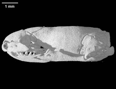
The skull of Casarea, which was described using high-resolution X-ray computed tomography by Masiano & Reippel (2007), is unique in having the maxilla subdivided into two movably jointed parts. That's right - the maxilla - the upper jaw. Snakes are renowned for the highly kinetic skulls, but no other snakes (or vertebrates, for that matter) have a kinetic maxilla. This jaw and its associated musculature are the basis for classifying Casarea and Bolyeria in a family of their own. Other lizard-eating snakes have analogous adaptations for grasping their hard-bodied prey, but no group takes this adaptation to such extremes as the bolyeriids. But think - on an island with no mammals and few birds, with little else but lizards to eat, selection is stronger than anywhere else for adaptations to saurophagy.

High-resolution X-ray computed tomography image of Casarea skull
Juvenile Casarea
Round Island keel-scaled boas reach 1 to 1.5 meters in length. Color changes ontogenetically (with age). According to observations made in captivity, Casarea are primarily nocturnal. Donald McAlpine, a researcher at the Jersey Wildlife Preservation Trust, where captive C. dussumeri are bred and studied, published a paper in 1983 showing data that captive specimens changed color every day, from light at night to dark during the day. Physiological color change in snakes has since been documented a few other times, such as in the boa constrictors of Cayos Cachinos, Honduras, and in the southeast Asian snake Enhydris gyii. Whether the purpose is cryptic, thermoregulatory, or something else entirely, we can only speculate. McAlpine ended his paper with the statement: "Hopefully this interesting phenomenon will be examined before Casarea becomes extinct." McAlpine's paper has been cited only twice, and as far as I can tell no research on this topic has been done since. An expedition to look for Bolyeria in 2001 was unsuccessful.

Painting of Bolyeria - no photos of living animals are known
ACKNOWLEDGMENTS


REFERENCES

Bauer A, Günther R, 2004. On a newly identified specimen of the extinct bolyeriid snake Bolyeria multocarinata. Herpetozoa 17:179-181.

Cundall D, Irish FJ, 1989. The function of the intramaxillary joint in the Round Island boa, Casarea dussumieri. Journal of Zoology 217:569-598.

Frazzetta T, 1971. Notes upon the jaw musculature of the Bolyerine snake, Casarea dussumieri. Journal of  Herpetology 5:61-63.

Hallermann J, Glaw F, 2005. Evidence for oviparity in the extinct bolyeriid snake Bolyeria multocarinata (Boie, 1827). Herpetozoa 19:82-85.

Korsós Z, Trócsányi B, 2006. The enigmatic Round Island burrowing boa (Bolyeria multocarinata): survival in the wild remains unconfirmed. African Herp News 40:2-7.

Maisano JA, Rieppel O, 2007. The skull of the Round Island boa, Casarea dussumieri Schlegel, based on high-resolution X-ray computed tomography. Journal of Morphology 268:371-384.

McAlpine DF, 1983. Correlated physiological color change and activity patterns in an Indian Ocean Boa (Casarea dussumeri). Journal of Herpetology 17:198-201.

Dwarf pipesnakes

$
0
0

Anomochilus monticola
A contest to be the most enigmatic snake would be like a contest to be the least well-known president serving between Jackson and Lincoln (seriously, name even one). But the dwarf pipesnakes (Anomochilidae) make even vice president Elbridge Gerry look like Marilyn Monroe by comparison.

Ok, not literally
It's hard to overstate how obscure anomochilids are. A paper published in 2007 using morphological and molecular data to examine the phylogeny of snakes was unable to incorporate the anomochilids because no molecular data on these snakes was available. This seems unbelievable in a day and age when it takes only a day and $1000 to sequence an entire human coding genome. Of course, it isn't inadequacies of technology that have kept the genes of anomochilids from us - it's lack of anomochilids, specifically fresh anomochilid tissue samples. In all museums in all the world, the Anomochilidae are represented by fewer than 15 specimens. Bigfoot has been seen more times than that.

But never collected
The first anomochilid was collected by Max Weber in Sumatra and described  in 1890 by Theodorus Willem van Lithe de Jeude, a curator of the Leiden Museum in the Netherlands (not to be confused with Erland of the same name, of Running Man fame). It was named Anomalochilus weberi after its collector, who also edited the volume in which its description was published, and it was one of only three snakes illustrated therein, out of 50 species covered. The spelling of the genus was changed from Anomalochilus to Anomochilus by Berg in 1901, because the former was already in use for a beetle.

Anomochilus weberi on the left (1, 2, & 3), on the right (4, 5, & 6) is Asthenodipsas malaccana, a pareatid 

The second anomochilid specimen was collected in 1915 by Edward Jacobson, also in Sumatra, and again described by van Lithe de Jeude in a 1922 paper in the journal Zoologische Mededelingen, who assigned it to the same species as the first, A. weberi. These two Sumatran specimens, together with one from Borneo, are all that we know of Anomochilus weberi.

Anomochilus weberi line drawing from de Rooij's 1917 book
The Reptiles of the Indo-Australian Archipelago
The second species of anomochilid wasn't discovered until 1940, when its description was published by Malcolm Smith of the British Museum in the Annals and Magazine of Natural History, the same journal in which Alfred Russel Wallace's 1855 paper "On the Law which has Regulated the Introduction of New Species" pre-empted Darwin's theory by four years. Smith named this species Anomochilus leonardi, again after its collector, G.R. Leonard, who may be the only person ever to have collected two anomochilidsIt is today known from five specimens collected in peninsular Malaysia (including the first, or type, and second, or paratype, specimens), and one from the Sabah province of northern Borneo. The Sabah specimen was collected by Raymond Goh in 1981, but sat undescribed in the Sabah Museum until 1993.

Figure from Smith 1940
Fast forward to 2002, when a snake collected in 1993 in northern Borneo was described in a book by Rudolf Malkmus and colleagues called Amphibians and Reptiles of Mount Kinabalu (North Borneo). The authors of the book called the snake Cylindrophis ruffus, which is a pipesnake in the family Cylindrophiidae. Although their book included photos of C. ruffus, the snake they described was in fact a third species of anomochilid, which was described in 2008 by Indraneil Das and colleagues. They named it Anomochilus monticola, because it was found in a mountainous area, and published the only photos of a living anomochilid known (above, in black and white). Based on the three specimens they had, they noted that A. monticola was far larger than either A. weberi or A. leonardi, and that it also differed in scalation and pattern. Their paper includes a nice history of anomochild discoveries, after which I have based most of this article.

Skull of Anomochilus leonardi imaged using high-resolution X-ray computed tomography
From Digimorph.org

One group of intrepid researchers, led by David Gower at the Natural History Museum in London, recently tried to extract DNA from three Anomochilus leonardi, with mixed success. Two of these were preserved over 50 years prior, and no genes could be recovered from their tissues. From one specimen collected in 2003, partial sequences of 12S and 16S rRNA mitochondrial genes were amplified, for a total of 221 informative sites. In their trees, Anomochilus leonardi formed a clade with Cylindrophis maculatus, rendering the latter's family, Cylindrophiidae, paraphyletic. However, this conclusion is based on limited data, and this study was done before the discovery of A. monticola, from which fresh tissue could presumably be obtained.

As early as 1890, van Lithe de Jeude noticed the similarities and differences between Anomochilus and other basal alethinophidians. He remarked that it was similar to Anilius scytale, a primitive snake from the Amazon rain forest, in that both lacked a mental groove (a structure on the chin that allows the lower jaw to open widely), but that the scalation of the head was more similar to Cylindrophis than Anilius. Respectively, Anilius, Anomochilus, and Cylindrophis are known as the red, dwarf, and Asian pipesnakes, and they were once treated as a single family together with shield-tailed (uropeltid) and sunbeam snakes (look out for future articles!), though today these are referred to separate families. In outward appearance, all of these snakes are have glossy scales, a result of their scale microornamentation, and a pattern of yellow or white spots and a red tail band against a dark ground color. They have blunt tails and few specialized head scales, with mostly undifferentiated ventrals. These basal lineages have much to teach us about snake evolution, if we can find enough of them to learn from!

Scanning electron microscope photograph of ventral scale
microornamentation of Anomochilus leonardi: BMNH 1946.1.17.4

Despite all the mystery, we do know some intriguing things about anomochilids. Unlike some basal snakes, but like others, Anomochilus has no pelvic girdle vestiges. However, it does possess vestiges of pectoral girdle muscle, unusual among snakes, which lack any vestige of a pectoral girdle bone. In addition, Anomochilus is unique in having lost the left lung entirely, a structure which is vestigial, but still present, in most other snakes. Like other basal snakes, Anomochilus has only a few teeth - 3 in each upper jaw and 5 in each lower jaw. What they eat is a matter of pure speculation, as are most details about how they reproduce (one female specimen contained four eggs, so we know that they are probably all oviparous, with small clutch sizes). Most of these basal alethinophidians eat elongate vertebrates, such as caecilians, amphisbaenians, and other snakes, because they do not have sufficiently flexible skulls to consume the very large prey items eaten by the macrostomate snakes (boas, pythons, and caenophidians).

ACKNOWLEDGMENTS

I would like to thank exactly no photographers, because apparently no one has ever taken a picture of these things, except for Das et al., who thoughtfully published photos of the type specimen of Anomochilus monticola in 2008.

REFERENCES

Cundall D, Rossman DA (1993) Cephalic anatomy of the rare Indonesian snake Anomochilus weberi. Zoological Journal of the Linnean Society 109:235-273

Cundall D, Wallach V, Rossman DA (1993) The systematic relationships of the snake genus Anomochilus. Zoological Journal of the Linnean Society 109:275-299

Das I, Lakim M, Lim KKP, Hui TH (2008) New species of Anomochilus from Borneo (Squamata: Anomochilidae). Journal of Herpetology 42:584-591

de Rooij N (1917) The Reptiles of the Indo-Australian Archipelago. Il. Ophidia. E. J. Brill, Leiden. 334 pp. <link>

Gower D, Vidal N, Spinks J, McCarthy C (2005) The phylogenetic position of Anomochilidae (Reptilia: Serpentes): first evidence from DNA sequences. Journal of Zoological Systematics and Evolutionary Research 43:315-320

Greene HW (1997) Snakes: The Evolution of Mystery in Nature. University of California Press, Berkeley
<this is the single best book on snakes available - if you don't own it, shame on you>

Lee MSY, Hugall AF, Lawson R, Scanlon JD (2007) Phylogeny of snakes (Serpentes): combining morphological and molecular data in likelihood, Bayesian and parsimony analyses. Systematics and Biodiversity 5:371-389

Malkmus R, Manthey U, Vogel G, Hoffmann P, Kosuch J (2002) Amphibians and Reptiles of Mount Kinabalu (North Borneo). Gantner Verlag, Rugell. 424 pp. <link>

Smith MA (1940) A new snake of the genus Anomochilus from the Malay Peninsula. Annals and Magazine of Natural History, Series 11 6:447-449 <link>

Stuebing RB, Goh R (1993) A new record of Leonard's pipe snake, Anomochilus leonardi Smith (Serpentes: Uropeltidae: Cylindrophinae) from Sabah, northwestern Borneo. Raffles Bulletin of Zoology 42:311-314

Tsuihiji T, Kearney M, Rieppel O (2006) First report of a pectoral girdle muscle in snakes, with comments on the snake cervico-dorsal boundary. Copeia 2006:206-215

van Lidth de Jeude TW (1890) Reptilia from the Malay Archipelago. II. Ophidia. In: Weber M (ed) Zoologische Ergebnisse einer Reise in Niederlandischost-Indien, vol 1. E. J. Brill, Leiden, The Netherlands, pp 178-192; PL XV-XVI <link>

van Lidth de Jeude TW (1922) Snakes from Sumatra. Zoologische Mededelingen 6:239-253 <link>

Yaakob N (2003) A record of Anomochilus leonardi Smith, 1940 (Anomochilidae) from Peninsular Malaysia. Hamadryad 27:285-286

Identifying snake sheds, part II

$
0
0

Not long ago, I posted about some techniques I used to identify a couple of snake sheds that I found in  Florida. I didn't plan a second post, because snake sheds are rarely found intact, but this week in southern Utah I had the opportunity to identify another snake shed, this one in nearly perfect shape. I found it snaked through the grass pointing at the opening of a burrow with an opening about the size of a quarter. Surely the snake had gone down the burrow, which led underneath a rock that was too large to lift (or else I would have!).

Habitat from the area
Despite being relatively fresh, the shed had already dried out, because it was extremely windy where I found it, at the base of a dam at Quail Lake State Park. In extricating it from the grass, I tore it at the midbody, which luckily didn't impair my ability to identify it later on. Importantly, the head was in perfect shape. I cupped it in my hands during the long walk back to the car, to prevent it being torn or blown away by the strong wind. As a result, I didn't get a chance to actually look at it closely for about half an hour, during which time a slew of possibilities ran through my mind as to what it could be. I am new to the southwest, so many of the species here are still unfamiliar to me. Because of what appeared to be a blunt head, as well as the overall small size (about 10 inches in SVL and 11.5 inches in total length), I first thought of a blindsnake, something I have wanted to see for quite some time. If the shed proved to be a blindsnake, I was prepared to recruit some serious help in lifting that rock. However, a glance through my fingers revealed differentiated ventral scales, which are characteristic of advanced snakes. I ruled out Scolecophidia.

Utah Blindsnake, Leptotyphlops humilis
There were many other possibilities, because the southwestern corner of Utah is in the Mojave desert, home to many species of reptiles that are not found in the rest of Utah. Another possibility that crossed my mind was the Southwestern Black-headed Snake, Tantilla hobartsmithi. Like the blindsnake, this species is adapted for burrowing. It is named for esteemed herpetologist Hobart Smith, who was born in 1912 and continues to conduct research and publish papers on reptiles and amphibians today, at age 99, despite having retired twice, in 1968 and 1983. Having published more than 1,600 manuscripts, Smith is the most published herpetologist of all time. He has described 102 species of reptile and amphibian, ranking 13th among all biologists in this regard.

Southwestern Black-headed Snake, Tantilla hobartsmithi
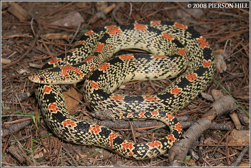
When the shed and I were safely in the car, however, I noticed that the head of my snake shed was not dark. Furthermore, the dorsal scales were boldly patterned with regularly-spaced dark blotches, twenty-eight in all (twenty-six on the body and two on the tail). The tail tip was broken, so I would guess that there were either thirty or thirty-one blotches in total. This was an important clue. The blotches were somewhat reminiscent of a kingsnake, milksnake, or long-nosed snake. However, they were restricted to the dorsal scales, rather than ringing the body as in king and milksnakes, and their edges were very clean, with no pattern in between, unlike the messier blotches of the long-nosed snake. Other options included the nightsnake and the glossy snake, but the blotches of my snake were very dark and regular, whereas these species have smaller, more irregular blotches.

Western Long-nosed Snake, Rhinocheilus lecontei
Finally, I turned to the scales for clues. As always, scale counts provide the most unambiguous evidence, although at this point I had a pretty good idea of what I thought it was. The dorsal scales were smooth and shiny, in 15 rows, and the subcaudal scales were divided, as was the anal plate. The head scales, most important, were somewhat reduced, consistent with a fossorial (burrowing) lifestyle. There were two postocular scales and only a single temporal scale in the first row, followed by two small temporals in the second row that I mistook for undifferentiated occipital scales at first. The upper labials were difficult to count, because the shed had already dried a little, and the snake had probably scraped it off using the labials as a leverage point. The same was true of the lower labials, but only a single pair of chin shields was evident.

Anterior part of the shed

Dorsal, lateral, and ventral views of the head

Rest of the body
After consulting some books to make sure I was right, I concluded that the shed belonged to a Ground Snake, Sonora semiannulata. These small snakes are highly variable in their body coloration and pattern, without consistent within-population variation. Although it is primarily restricted to the Mojave portion of Utah, records from the northeastern and central parts of the state suggest that it might be more widespread. It is found from southwestern Missouri west to southern California, north to Oregon and Idaho, and south to Mexico. Like other members of the tribi Sonorini, Ground Snakes eat mostly arthropods, including insects,  scorpions, spiders, and centipedes. Little is known about the species despite its wide range.

Ground Snake, Sonora semiannulata
It was exciting, almost forensic, to identify the shed of a species I had never seen before. Now I had a debate on my hands about whether to include it on my life list or not (a life list is a compilation of all the species of something - often birds, but in my case herps - that an individual has seen in their life). My friend Kerry Nelson and I have had lengthy discussions about what counts and what doesn't, including whether animals that others find are valid, whether dead animals are valid, and whether or not species seen in dreams (including those that exist only in dreams) are valid. What do you think? Would you count a shed, unambiguously identified, as seeing a species? I decided against it, but I'm very much looking forward to finding a live ground snake so I can add it to the list!

ACKNOWLEDGMENTS

I would like to thank Brian EagerMatthijs Hollanders, Pierson Hill, and William Flaxington for use of their photographs.

REFERENCES

Cox DT, WW (1995) Snakes of Utah. Bean Life Science Museum, Provo, UT

Ernst CH, Ernst EM (2003) Snakes of the United States and Canada. Smithsonian Institution Press, Washington D.C.

Uetz P (2010) The original descriptions of reptiles. Zootaxa 2334:59-68 <link>

The snakes that eat caviar

$
0
0


Banded sea krait, Laticauda colubrina
Marine snakes are fascinating. Entire articles have been written about their morphological and physiological adaptations to marine life, from their lingual salt glands, which are more efficient than kidneys at removing sodium ions from their body, to their rudimentary left lung, which serves a function for the first time in millions of years, aiding in buoyancy control in a manner analogous to the swim bladders of many fishes. There appear to have been three separate invasions of the ocean by terrestrial snakes, all from the family Elapidae, which also includes cobras, mambas, and coral snakes. Although they have spread to east Africa and the south Pacific, all of these invasions have taken place in the shallow seas around Australia and southeast Asia. This is the center of elapid species diversity, so it's no surprise that the greatest ecological diversity is also found here. Among the nearly 70 species of sea snake, however, two genera in particular stand out.

Most marine snakes eat eels and other tropical shore fishes, in accordance with their ancestors' diets of large, bulky prey items that required venom or constriction to subdue. But in 1966, Harold Voris reported that the stomachs of two species of sea snake in the genus Emydocephalus, the turtle-headed sea snakes, were found to contain only fish eggs. This was a remarkable discovery, because most snakes eat prey that are relatively large compared to themselves, and they do so infrequently. It's perhaps one of the evolutionary novelties that has allowed snakes to be so successful. But Emydocephalus eats tiny eggs, and it does so several times an hour, using a foraging mode similar to herbivorous browsing mammals, and to the lizard ancestors of snakes, than to other snakes. Turtle-headed sea snakes use chemoreception to locate the eggs, and the parent fishes are sometimes able to chase them away, despite being far smaller. The parent fishes are never eaten, and indeed they have little to fear, except for their fitness. Why?

There's a reason there was no turtle-headed sea snake character in Finding Nemo
Voris also noticed that the dentition of these snakes was highly unusual, in that they almost completely lack teeth. Most snakes have teeth on up to five of their skull bones on each side: the maxilla, premaxilla, palatine, pterygoid, and dentary. Three of these bones (maxilla, premaxilla, and dentary) also bear teeth in humans and other mammals - the first two in your upper jaw, and the dentary (also called the mandible) in the lower. The palatine and pterygoid teeth of snakes are located on the bones that form the roof of your mouth, and they form what is essentially a second set of upper jaws inside of the first, which can move independently of the outer upper jaws and of each other. In Emydocephalus, only the pterygoid bone has any teeth, except for a single large proteroglyphous fang on each maxilla.

Partial skull of three species of sea snake, looking at the roof of the mouth from below.
Figure modified from McCarthy 1987
 
It's clear that a snake that ate only soft fish eggs wouldn't need those teeth, but Voris couldn't figure out how Emydocephalus actually ate fish eggs. He did notice that their stomachs also contained a good bit of sand, and occasionally a copepod (a type of crustacean). In 1987, Colin McCarthy proposed a mechanism that is very similar to that used by most fishes: suction. Based on his observations of the throat musculature of a closely related sea snake, Aipysurus eydouxi, also known to eat fish eggs, he suggested that the two genera of egg-eating sea snakes could create suction by contraction of the geniomucosalis muscle, which originates on the lower jaw and inserts on the oral mucosa (the lining of the mouth). The same mechanism is used by blindsnakes (Scolecophidia), the taxon in which the muscle was described only eight years earlier, to create suction as they feed on ant and termite pupae and larvae.

Graph showing the number of true sea snakes that feed on a variety of prey shapes
From Voris and Voris, 1983
Other modifications of the head aid Emydocephalus and Aipysurus in finding and consuming fish eggs. Most snakes have six to eight labial scales (scales along the lip), whereas Emydocephalus has only three, giving it the appearance of a beak similar to that of a turtle (its genus name means 'turtle-headed' in Greek). McCarthy thought this helped keep the lips rigid during suction feeding. A spine at the tip of the rostral scale might aid in probing the sand for fish eggs buried there, but a secondary sexual function is also likely, because only adult male Emydocephalus have it.

Male Emydocephalus annulatus
In 1996, Michael Guinea published some of the first behavioral observations of wild Emydocephalus from northwestern Australia. While snorkeling, he watched as many as twenty individual E.annulatus interact on a circular coral mass only 25 feet in diameter. Algae grew on them, they moved so little. Mating males touched females with their spines, which might help them synchronize hourly trips to the surface for air and keep track of the female's location as the pair return to the bottom, where Guinea observed pairs mating for over an hour. He also observed E. annulatus using their enlarged labial scales to scrape damselfish eggs off coral, but did not notice any evidence of suction feeding. He suggested that the geniomucosalis muscle was  instead used in rapid exhalation at the surface, and noted that exhalations of Emydocephalus can be heard, whereas those of other sea snakes lacking a geniomucosalis cannot (unlike Emydocephalus, other sea snakes exhale on their way to the surface, leaving a trail of bubbles).

Emydocephalus annulatus courting
You might have immediately associated sea snakes with potent venom, and you're right to do so. It has been suggested that these marine snakes evolved simple, especially fast-acting venoms to immobilize their fish prey, which can escape in three dimensions rather than just two. However, Min Li and colleagues examined the venom of Aipysurus eydouxii and found a mutation that caused a 50- to 100-fold decrease in venom neurotoxicity. They also noted that A. eydouxii has greatly atrophied venom glands and relatively ineffective fangs. In their words, "It is interesting to note that a potent venom was not maintained for use in defense, thus reinforcing that the primary use of snake venom is for prey capture." This is the first case of decelerated
evolution of toxins in snake venom, which is usually evolving rapidly, in an "arms race" with the immune system of the prey. Emydocephalus also have reduced fangs and venom glands, but no study of the chemical properties of their venom has been undertaken.

Aipysurus eydouxii
Are there any freshwater snakes that have similar adaptations to  Emydocephalus and Aipysurus? There are plenty that fill similar ecological roles to other sea snakes, eating fishes and crustaceans. There are lots of fishes and amphibians that lay tasty eggs in fresh water, but no freshwater snakes are known to have anything close to the morphological adaptations for oophagy of  Emydocephalus and Aipysurus. There are some terrestrial snakes that eat eggs, such as the neotropical Leptoderia, the African Dasypeltis, and the Australian Brachyurophis, the latter two of which  have lost many of their teeth and are incapable of eating other prey.

Leptodeira annulata eating Agalychnis callidryas eggs 

ACKNOWLEDGMENTS

Thanks to the Field Museum archive for many of these images, and to Klaus Stiefel and il_mare77.

REFERENCES

Guinea ML (1996) Functions of the cephalic scales of the sea snake Emydocephalusannulatus. Journal of Herpetology 30:126-128

Li M, Fry B, Kini RM (2005) Eggs-only diet: its implications for the toxin profile changes and ecology of the marbled sea snake (Aipysurus eydouxii). Journal of Molecular Evolution 60:81-89 <link>

Li M, Fry BG, Kini RM (2005) Putting the brakes on snake venom evolution: the unique molecular evolutionary patterns of Aipysurus eydouxii (Marbled sea snake) phospholipase A2 toxins. Molecular Biology and Evolution 22:934-941

McCarthy C (1987) Adaptations of sea snakes that eat fish eggs; with a note on the throat musculature of Aipysurus eydouxi (Gray, 1849). Journal of Natural History 21:1119-1128

Shine R, Bonnet X, Elphick M, Barrott E (2004) A novel foraging mode in snakes: browsing by the sea snake Emydocephalus annulatus (Serpentes, Hydrophiidae). Functional Ecology 18:16-24 <link>

Voris HK (1966) Fish eggs as the apparent sole food item for a genus of sea snake, Emydocephalus (Krefft). Ecology 47:152-154 <link>

Voris HK, Voris HH (1983) Feeding strategies in marine snakes: an analysis of evolutionary, morphological, behavioral and ecological relationships. American Zoologist 23:411-425

Snake-eating beetles

$
0
0

So little is known about the parasites of snakes that we tend to discount them all together, but the ecological  and evolutionary interactions between hosts and their parasites can be very strong. This is a story about how two enterprising snake biologists solved a mystery that had been puzzling entomologists for decades.

Burying beetles (genus Nicrophorus) conceal small vertebrate carcasses underground and prepare them for consumption by their young by excavating a crypt up to 60 cm deep, removing fur or feathers from the carcass, and covering it in anal secretions to prevent fungal growth. The two parents slowly eat the carcass, defending it from other carrion eaters, and feed regurgitated bits of it to their altricial larvae, which hatch from eggs they lay in the walls of the crypt and beg to be fed like baby birds. Although feeding your babies poop-coated vomit sounds like the plot of a gruesome horror movie, it has been a successful evolutionary strategy for the burying beetles. Their complex social behavior, including biparental care and communal breeding, is unusual among insects. The whole process takes about two weeks.

Nicrophorus pustulatus
Of the nearly 75 species of burying beetle, distributed throughout the Northern Hemisphere, one in particular stands out for its unusual natural history. Entomologists studying the group use dead mice to bait traps, but one species, Nicrophorus pustulatus, never seemed attracted to the carrion. In addition, they are able to produce very large broods (up to 190 vs. 30-45 for most other species of burying beetle) of large offspring on carcasses in the laboratory. Usually, a large brood size comes hand-in-hand with a decrease in individual offspring size, but not in this species apparently. Why not?

Theories ranged from that N. pustulatus used larger carcasses, such as rabbits, without burying them, to that  it was an interspecific brood parasite, like a brown-headed cowbird, laying its eggs in the nests of other burying beetles. But in 2000, a paper in the journal Ecoscience by two snake biologists, Gabriel Blouin-Demers and Patrick Weatherhead, then of Carleton University in Ontario, revealed a surprising discovery. They were studying the nesting ecology of black ratsnakes (Pantherophis obsoletus, formerly Elaphe obsoleta) in Canada by radio-tracking adult female ratsnakes to their oviposition sites. Their purpose was to document the use of communal nests by these snakes and to collect information on clutch size and juvenile survival. When they examined the ratsnake nests they found, they discovered that many of them contained adult and larval  N. pustulatus.

Ratsnake eggs parasitized by carrion beetles.
Blouin-Demers and Weatherhead found evidence of beetles in six of the seven nests they looked at. In some nests, only old eggshells with small holes evidenced the beetles' presence, but in others 100% of the eggs were destroyed by the beetles and their larvae. Because black ratsnakes nest communally in this part of the world, up to 111 eggs can constitute a nest, even though the average clutch size is only 11-15 eggs per female. The ratsnakes use the same communal nesting sites year after year, which can be highly beneficial because of increased nest temperature and shorter development time. At such northern latitudes, female ratsnakes do not lay eggs until June or July, and the babies must hatch by late August in order to avoid being killed by an early frost. A mother ratsnake's only parental care is her nest site choice, and research has shown that eggs laid in communal nests hatch earlier, grow larger in their first year, and can even swim faster than those incubated with just their clutchmates. However, the probability of a beetle infection probably increases with increasing nest size, because it only takes one infected egg to spread the beetles to the whole nest. This is why N. pustulatus is so fecund compared to other carrion beetles - because it can raise enormous numbers of larvae on large snake nests, full of nutritious eggs and already hidden away in sites with ideal thermal and humidity.

Black Ratsnake (Pantherophis obsoletus)
Based on their findings, Blouin-Demers and Weatherhead characterized N. pustulatus as a parasitoid of snakes. A parasitoid is different from a parasite because they are parasitic only as larvae (although in this case, with a little help from their parents), and they always kill their host. However, they are also different from predators, because each parasitoid larva only kills a single host individual instead of many. Blouin-Demers and Weatherhead suggested that theirs was the first example of a vertebrate being host to an arthropod parasitoid, and so far they are correct.

The full mystery is far from solved, though. Did N. pustulatus evolve this behavior by first exploiting snake eggs that failed to hatch? How do the beetles find reptile eggs? Are communal nests easier for the beetles to find, or do they simply prefer them because of their higher concentration of resources? How has parasitism by this beetle influenced ratsnake evolution? Do any other species of Nircophorus also parasitize reptile eggs? Does N. pustulatus beetles also parasitize the eggs of other species of snake? Observations of fox snake (Pantherophis vulpinus) nests in Illinois have also yielded beetle larvae. The range of N. pustulatus extends farther north than that of any oviparous snake species (snakes at high latitudes tend to be viviparous, because the females can more precisely control the temperature of their developing offspring if they carry them around). What do they use for rearing their young up there? Could it be turtle eggs, or do they use small animal carcasses like their ancestors?

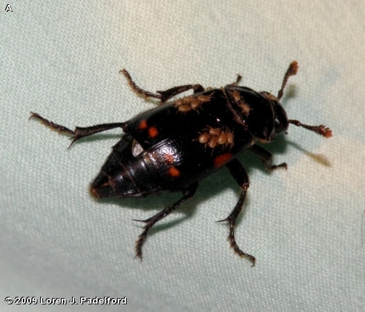
Nicrophorus pustulatus with phoretic mites
From the beetle's perspective, it has arrived at a very successful reproductive strategy by shifting hosts. By moving away from nesting in carcasses, for which they must compete with flies, ants, fungi, bacteria, and scavenging vertebrates such as skunks and raccoons, it has secured an apparently unique niche. As a defense against carcass competitors, some Nicrophorus species carry phoretic mites that eat fly eggs, but lab experiments have shown that the mites sometimes eat the beetles' eggs too, so the benefit is not without risk. Additionally, not having to move or bury snake eggs saves the parent beetles a lot of energy prior to laying their eggs. Experiments have shown that N. pustulatus females oviposit rapidly in house snake (Lamprophis) eggs, and that male beetles elevate their sex pheromone emission in response to snake eggs. Other beetles in the genus Nicrophorus did not show the same response. While N. pustulatus will use mouse carcasses to rear their young in the lab, no one has ever found them doing so in the field. The entomologists who performed these lab tests also found that N. pustulatus adjusted its fecundity to the available mass of snake eggs.

As a driver of evolution in oviparous snake nesting strategies, Nicrophorus pustulatus may play an important role. Could they potentially pose a threat to egg-laying snake species that are of conservation concern, such as the Eastern Indigo Snake (Drymarchon couperi)? What might happen if they were introduced to a continent whose snakes had not evolved with parasitic beetles eating their eggs? There is still so much we don't understand about snake behavior, reproduction, ecology, and evolution, especially in the wild. Thanks to the observations of a few scientists who thought they were studying something else entirely, we are one step closer.


ACKNOWLEDGMENTS

Thanks to photographers Joyce Gross, Loren Padelford, and Gabriel Blouin-Demers.

REFERENCES

Blouin-Demers G, Weatherhead PJ (2000) A novel association between a beetle and a snake: parasitism of Elaphe obsoleta by Nicrophorus pustulatus. Ecoscience 7:395-397


Blouin-Demers G, Weatherhead PJ, Row JR (2004) Phenotypic consequences of nest-site selection in black rat snakes (Elaphe obsoleta). Canadian Journal of Zoology 82:449-456


Ikeda H, Kubota K, Kagaya T, Abe T (2006) Niche differentiation of burying beetles (Coleoptera: Silphidae: Nicrophorinae) in carcass use in relation to body size: estimation from stable isotope analysis. Applied Entomology and Zoology 41:561-564

Robertson IC (1992) Relative abundance of Nicrophorus pustulatus (Coleoptera: Silphidae) in a burying beetle community, with notes on its reproductive behavior. Psyche 99:189-198

Scott MP (1998) The ecology and behavior of burying beetles. Annual Review of Entomology 43:595-618

Smith G, Trumbo S, Sikes D, Scott M, Smith R (2007) Host shift by the burying beetle, Nicrophorus pustulatus, a parasitoid of snake eggs. Journal of Evolutionary Biology 20:2389-2399

Trumbo ST (2007) Defending young biparentally: female risk-taking with and without a male in the burying beetle, Nicrophorus pustulatus. Behavioral Ecology and Sociobiology 61:1717-1723

Snakes that chew their food

$
0
0

I have to admit right up front that the title of this article is not really accurate. No snakes chew their food the way we do. Almost all snakes must swallow their food whole, which limits their (often considerable) gape to items they can jaw-walk their kinetic skulls over. Taken as a whole, there are few animals on Earth that snakes do not eat -- whales and dolphins, elephants, animals endemic to the polar regions, some very toxic millipedes. There are snakes that swallow leopards whole, snakes that eat porcupines without removing the quills, snakes that tolerate stabs from catfish spines, snakes that eat other snakes longer than they are. Here's a video of a Tantilla eating a giant centipede. As a group, they can eat nearly anything. They all swallow their prey whole. Almost.
Except for this one
Its genus name is Fordonia, which is probably meaningless, seeing as it was biologist J.E. Gray of the British Museum of Natural History,"well known for inventing many apparently meaningless scientific names", who came up with it (he also named the North American Farancia). Commonly known as the Crab-eating Water Snake or White-bellied Mangrove Snake (after the specific epithet), Fordonia leucobalia is native to the mangrove swamps and tidal mud flats of southeast Asia and northern Australia. It lives in mud lobster and fiddler crab burrows, and moves by jumping across the soft mud, into which it would sink if it tried to slither.

Part of a small but interesting group of live-bearing snakes known as homalopsids, Fordonia is southeast Asia's answer to the North American natricine Nerodia, for many the archetypical semi-aquatic snake. What sets Fordonia apart from other homalopsid snakes, which feed mostly on fishes, is that it eats crabs, an observation first made by Cantor in 1847. (This may be highly cathartic for the snakes, whose primary predators as juveniles include large crabs.)

Those are hard-shelled decapod crustaceans, for you biologists out there 
Like many other arthropods, crabs have an anti-predator adaptation called leg autotomy, similar to tail autotomy in lizards, salamanders, and some snakes. This means that their legs can break off when grabbed and will later regrow - better to lose a limb and escape than to be eaten whole. But Fordonia has evolved behaviors that exploit the crabs' ability to autotomize their legs - it pins the crab's body to the mud and pulls of its legs, eating them one at a time! Sometimes they also consume the crab's body, but often they just leave it behind. This makes Fordonia the only snake that breaks its prey apart prior to eating it, although we must admit that it is somewhat helped along by the crab's autotomy. This discovery was sufficiently exciting to be published in the prestigious journal Nature.

The five crab legs at the top, eaten by this snake, came from a crab about the size of the one on the bottom. The white circle represents the maximum-sized prey item the snake could have eaten whole. Figure from Jayne et al. 2002
The adaptations of Fordonia to cancrivory don't end there. As anyone who has eaten crab legs knows, a crab's exoskeleton is very tough - we humans must use tools to break into it. In order not to be internally lacerated by their prey, Fordonia have evolved extra tough, muscular stomach lining. Other crustacean-eating snakes, such as the North American crayfish snakes (genus Regina), as well as the arthropod-eating False Hook-nosed Snake (Pseudoficimia frontalis, a sonorine snake from western Mexican dry forests), also have thickened muscles surrounding their stomachs, to prevent internal damage from they prey's sharp exoskeletons.


Digestion in snakes is an intense process: their digestive enzymes are very strong, capable of breaking down  even bone. Still, a little mastication can help the digestive process along considerably. For most snakes this isn't an option, because their needle-like teeth and highly mobile skull bones are ill-suited to both cutting and generating bite forces. However, snake biologist Alan Savitzky reported that recently ingested crab legs extracted from Fordonia stomachs were crushed. How is this possible? In fact, Fordonia possess remarkably robust and compact teeth for a snake, almost like molars! Although this is an extreme morphological modification, Savitzky remarked that it is almost surprising that the teeth and skulls of Fordonia aren't more abnormal, considering their unusual diet. Finally, Fordonia has evolved a large salt gland to help maintain osmotic balance on a high-salt diet (crabs are isosmotic to their environment, meaning that they have the same salt content as sea water).

Left: Tooth of Cerberus rynchops; Right: Teeth of Fordonia leucobalia
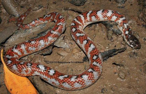
While Fordonia does all this with hard-shelled crabs, another homalopsid species found in the same mangroves, the Cat-eyed Watersnake (Gerarda prevostiana), has been found to consume freshly-molted (and therefore soft-shelled) crabs in much the same way. This kind of specialization is also found among the four species of North American Crayfish Snakes (Regina) - two of which (R. rigida, R. alleni) have hinged teeth to help them consume hard-shelled crayfish, and two of which (R. grahamii, R. septemvittata) seek out freshly-molted crayfish by smelling their molting secretions. Incredibly, although Gerarda lacks the morphological adaptations for cancrivory of Fordonia, it was observed tearing apart the soft carapaces of crabs after eating their legs, which probably allows Gerarda to consume crabs that would otherwise be too large for them to swallow whole. The feeding mechanisms used by Fordonia and Gerarda differ in the modes of attack and prey restraint, the usual orientation for swallowing crabs, and how pieces were torn from prey, suggesting that they might have evolved their crab-eating habits independently and convergently, rather than inheriting them from a shared common ancestor (although they are evolutionary sisters, one another's closest relatives). Two other closely related genera of homalopsine, Myron and Cantoria, also consume some crustaceans, but are less well-known. How many snakes are out there with strange dietary adaptations that remain to be discovered? We may never know.

This snake only eats soft-shelled crabs - what a snob

ACKNOWLEDGMENTS

Thanks to A. Captain and Brendan Schembri for photographs.

REFERENCES

Alfaro ME, Karns DR, Voris HK, Brock CD, Stuart BL (2008) Phylogeny, evolutionary history, and biogeography of Oriental-Australian rear-fanged water snakes (Colubroidea: Homalopsidae) inferred from mitochondrial and nuclear DNA sequences. Molecular phylogenetics and evolution 46:576-593

Jayne BC, Voris HK, Ng PKL (2002) Snake circumvents constraints on prey size. Nature 418:143

Savitzky AH (1983) Coadapted character complexes among snakes: fossoriality, piscivory, and durophagy. American Zoologist 23:397-409

Shine R, Schwaner T (1985) Prey constriction by venomous snakes: a review, and new data on Australian species. Copeia 1985:1067-1071

Voris HK, Jeffries WB (1995) Predation on marine snakes: a case for decapods supported by new observations from Thailand. Journal of Tropical Ecology 11:569-576

Voris HK, Murphy JC (2002) The prey and predators of Homalopsine snakes. Journal of Natural History 36:1621-1632

Egg-eating snakes

$
0
0

I think we can all agree that amniotic eggs are delicious. They also happen to be one of the best sources of energy out there, and this is at least partially why we, and many other animals, enjoy eating them so much. In addition, they rarely fight back, and they almost never have physical defenses, such as spines, or chemical ones, such as deadly toxins. In fact, on the inside they're pretty much all lipids (a group of molecules including fats and cholesterol), surrounded by either a leathery (in monotremes and most reptiles) or a hard, calcified (in birds) shell. I've already written about a species of burying beetle that specializes on snake eggs, apparently with great benefit to its fecundity relative to other burying beetles that use carrion. Turns out, snakes aren't above specialized oophagy themselves.

There are a few snakes that eat anamniotic eggs, such as the turtle-headed sea snakes (about which I've written before) and the South American goo-eaters. These have many amazing adaptations to eating shell-less eggs, but I'd like to focus on the amniotic egg-eating snakes for now. To review, an amniotic egg is one with a shell and several other embryonic membranes, called the amnion, chorion, and allantois. These structures physically protect the embryo and facilitate gas and waste exchange between the embryo and its surroundings, because the shell is too thick to allow the embryo to breathe and excrete by diffusion alone. These eggs are laid by birds, many reptiles, and monotremes (egg-laying mammals such as the platypus and echidna). In placental mammals (including humans), which are also amniotes, some of these structures are part of the umbilical cord, while others are vestigial. Amniotic eggs are adapted for being laid on land, and even the most aquatic of amniotes, such as sea turtles and pelagic birds, must come to land to lay their eggs.

Because of the resilience and self-contained nature of amniotic eggs, many organisms that lay them have done away with parental care. Choosing a nest site, usually under a rock, log, or pile of poop, or in a nest dug underground, is the extent of it. Beyond that, a female snake or turtle will most likely never see her kids hatch, let alone grow up, graduate, or become successful. This also means that their eggs are basically undefended from predators, except for being concealed and not smelling very much. Birds are slightly better parents, but they risk giving away the location of their nest to predators by flying back and forth to it many times a day. Experiments conducted by herpetologist Steve Mullin and ornithologist Bob Cooper have shown that gray ratsnakes locate bird nests over twice as quickly when parents are attending than when they aren't, a phenomenon so prevalent that it has its own name (Skutch's hypothesis) and is thought to influence the evolution of optimal clutch size in birds (because more offspring need to be fed more often, necessitating more trips to and from the nest and increasing the likelihood of detection by a predator).

Ok, enough - let's get to the pictures of snakes eating eggs!

African Egg-eating Snake, Dasypeltis scabra
Holy shit, how do they do that!? That snake is going to choke itself! Got to be a faked, Photoshopped image, right? Think again:


Damn, that's impressive. If you watched the video above, you saw an African Egg-eating Snake, perhaps the most specialized oophagous snake there is, swallow a bird egg whole, crack it open, and regurgitate the  shell. How does it do it? The highly kinetic, flexible skull of this snake allows it to maneuver its jaws around an egg many times bigger than its head, despite the smooth, round surface and the snake's lack of hands. It'd be like a human trying to eat a whole watermelon. Egg-eating snakes lack teeth almost entirely, not needing them for gripping their prey. In addition, the snake's skin is stretchy enough to accommodate the egg's passage - the scale rows are clearly visible, widely separated by the skin in between. Most of the time, this skin can't be seen, because the skin is relaxed so that the rows of scales are in contact with one another.

Once the egg is in the snake's esophagus, how does it get cracked open? Snakes have strong digestive juices, but waiting for them to dissolve the shell of an egg would take too long. OK, are you ready? This is the coolest part:

Vertebral hypapophyses of  African egg-eating snakes, Dasypeltis
See those spines? Those are called hypapophyses, which is a fancy term for things that stick off the bottom (ventral side) of vertebrae. You've got them too - but in egg-eating snakes, they're modified to be much larger and sharper, the better to pierce eggshells with, my dear. At least, the ones on vertebrae 17-38 are, the vertebrae that sit right above the esophagus and thus above egg once it has been swallowed. The esophagus itself is modified as well - it has loose folds, like pockets, into which each of the hypapophyses fits, so that they don't puncture the esophagus itself. See how it works in the following video, from the BBC's Life in Cold Blood:


Starting at 2:45, you can see the moving x-ray of the egg-eating snake swallowing the egg. Continuing through the end of the video, the snake cracks the shell, allows the yolk inside to drain into its stomach, and regurgitates the eggshell. Most amazing, young Dasypeltis don't appear to have these hypapophyses - they grow as the snakes get older, which raises questions about what the juveniles eat. Even though eggs are nutritious, Dasypeltis must feed relatively often for a snake - one that my advisor kept in captivity ate several quail eggs a week.

Lateral view of the skull of Dasypeltis, from Gans 1952
The adaptations of the nine species of Dasypeltis allow them to eat eggs that are very large relative to their body size, and as far as we know they eat almost nothing else. Several generalist snakes also eat eggs; adult Eastern Kingsnakes (Lampropeltis getula), Western Hog-nosed Snakes (Heterodon nasicus), and Formosa Kukrisnakes (Oligodon formosanus) frequently consume reptile eggs, and many members of the rat snake genera Pantherophis and Elaphe opportunistically feed on both eggs and nestling birds. These snakes, however, have no special morphological or behavioral adaptations to assist them in the consumption of eggs. One species, the Japanese rat snake (Elaphe climacophora), can ingest relatively large eggs, and has several vertebral hypapophyses. However, E. climacophora ingests the entire egg, including the shell. Only Dasypeltis, and possibly a poorly-known species from India called Elachistodon westermanni, specialize in ingesting large eggs, then crushing the shell and retaining solely the contents.

Defensive display by Dasypeltis scabra
ACKNOWLEDGMENTS

Thanks to Armata, Tony Phelps, and the BBC for images and videos.

REFERENCES

Coleman K, Rothfuss LA, Ota H, Kardong KV (1993) Kinematics of egg-eating by the specialized Taiwan snake Oligodon formosanus (Colubridae). Journal of Herpetology 27:320-327

Gans C (1952) The functional morphology of the egg-eating adaptations in the snake genus Dasypeltis. Zoologica 37:209-244

Gans C, Oshima M (1952) Adaptations for egg eating in the snake Elaphe climacophora (Boie). American Museum Novitates 1571:1-16

Gartner G, Greene H (2008) Adaptation in the African egg-eating snake: a comparative approach to a classic study in evolutionary functional morphology. Journal of Zoology 275:368-374

Mullin SJ (1996) Adaptations facilitating facultative oophagy in the gray rat snake, Elaphe obsoleta spiloides. Amphibia-Reptilia 17:387-394

Mullin SJ, Cooper RJ (1998) The foraging ecology of the Gray Rat Snake (Elaphe obsoleta spiloides)—visual stimuli facilitate location of arboreal prey. The American Midland Naturalist 140:397-401

Savitzky AH (1983) Coadapted character complexes among snakes: fossoriality, piscivory, and durophagy. American Zoologist 23:397-409

This blog is supposed to be about snakes, but if you can't make exceptions for family, then you're a jerk

$
0
0

Sphaerodactylus elegans of Cuba
Geckos are some of the most diverse and widespread squamates on Earth. They range in size from the dwarf Sphaerodacylus of the Antilles (16-18 mm) to the (probably) extinct Kawekaweau or Delcourt's Giant Gecko, Hoplodactylus delcourti, of New Zealand (as long as a tuatara and as thick as a man's wrist). Except for a few species, none have eyelids, and most have adhesive pads on their toes that allow them to climb slick surfaces. Some are parthenogenic. A few owe their success partly to humans, and it is these that we consider here today.

Several species of gecko have been introduced to the United States by humans, most accidentally, as stowaways or through the pet and greenhouse trade. One of the most ubiquitous is the Mediterranean House Gecko, Hemidactylus turcicus. Described by Linnaeus in the first edition of his Systema Naturae, it is a yellow-tan, nocturnal, insectivorous gecko about six inches long. Whereas most of our non-native lizards are limited to a few small areas in Florida, H. turcicus can be found in 17 states, from California to Maryland. It isn't continuously distributed across the southern US, but rather locally common in urban sites, due to many separate introductions, the earliest of which occurred in Key West before 1915. One such population is in a middle school in Cary, North Carolina.

Hemidactylus turcicus
Legend has it that a science teacher during the early 1980s was keeping some H. turcicus in a terrarium. All was well until an absentminded student left the lid off one day. The lizards escaped, and a small population has been living in the walls of the school campus ever since. At least, that's what everyone thought. An ongoing summer of research conducted by a wildlife student at North Carolina State University, my brother Kevin Durso, in 2012, has revealed that the population is much larger than anyone thought.


Bags of geckos
On the first night, Kevin counted 82 geckos on the walls of the campus buildings. He came back with reinforcements - tall college students and volunteers armed with lizard nooses and water guns, for blasting geckos off ceilings and walls. On some nights, students at the middle school come out with their teachers and parents to see what the research is all about. Kevin & Co. are marking each gecko they capture with glow-in-the-dark elastomer, so they can be individually identified upon later capture. Once captures of new, unmarked geckos begin to decline, he can begin to estimate the total size of the population using a mathematical model. Whether this will happen sooner or later is still hard to know. Kevin is also keeping track of the exact location of each gecko sighting, so that a home range size estimate can be made. He works at night, when the geckos are most active. Perhaps this is why no one knew the true size of the gecko population until now - how often is anyone at school at night? Not if I can help it, Mom!

Super Soaking a gecko off a wall

Previous research on house geckos has revealed that they inhabit similar areas, both climatically and in terms of microhabitat, in their native and non-native range. A population on the Stephen F. Austin State University campus in Nacogdoches, Texas, ate mostly grasshoppers, moths, and isopods. Their great success in southern North America has been attributed to low predation pressure, little interspecific competition, and a life history which maximizes survival at all ages. House geckos in southern Louisiana are host to native North American parasitic worms, so there is some potential for parasites to regulate populations of these non-native lizards.

Injecting a gecko with glow-in-the-dark elastomer
What factors have allowed the Cary H. turcicus population to grow so large? What effects do these non-native geckos have on the local ecosystem, from the arthropods they eat to the birds and snakes they are eaten by? Have they been spreading around the Triangle area since the 1980s, brought home on schoolbuses in students' backpacks and coats? Only time, and further research, will tell.


ACKNOWLEDGMENTS

The College of Natural Resources and the Office of Undergraduate Research at NC State University provided support and funding for this project. Thanks to Konrad Mebert, Miguel Landastoy, Alex Morrison, Kevin Durso, and Sandy Durso for photographs.

REFERENCES

Davis WK (1974) The Mediterranean gecko, Hemidactylus turcicus in Texas. Journal of Herpetology 8:77-80

Rödder D, Lötters S (2009) Niche shift versus niche conservatism? Climatic characteristics of the native and invasive ranges of the Mediterranean house gecko (Hemidactylus turcicus). Global Ecology and Biogeography 18:674-687

Rose FL, Barbour CD (1968) Ecology and reproductive cycles of the introduced gecko, Hemidactylus turcicus, in the southern United States. American Midland Naturalist 79:159-168

Saenz D (1996) Dietary overview of Hemidactylus turcicus with possible implications of food partitioning. Journal of Herpetology 30:461-466

Selcer KW (1986) Life history of a successful colonizer: the Mediterranean gecko, Hemidactylus turcicus, in southern Texas. Copeia 1986:956-962

Snake-eating snakes

$
0
0

I've mentioned before how snakes can eat nearly anything, due to amazing adaptations of their head and jaws that allow them to swallow objects bigger than their heads. But surely they must be limited to eating prey that are shorter than they are in overall length, right? What about items that are just a bit longer?

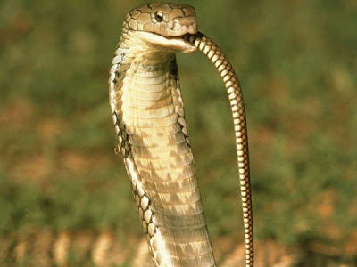
There are a wide variety of snakes that eat other elongate vertebrates, including other snakes, legless lizards, lizards with relatively small legs (like skinks), amphisbaenians, caecilians, and eels (today we'll focus on snakes, but look out for future articles on some of these other specialized diets). In many ways, this kind of diet is convenient for a snake, because they are already elongate, so they don't have to deform their stomachs, bodies, and mouths to the same extent as snakes that eat bulkier prey. Many are relatively primitive snakes that retain the robust skulls of their lizard-like ancestors, but quite a few derived snakes are snake-eaters as well, including the King Cobra, Ophiophagus hannah, the world's largest venomous snake.

The genus Ophiophagus means 'snake-eating'

Other familiar snake-eaters include the North American Kingsnakes (genus Lampropeltis), which have evolved resistance to the venom of many species of viper. Eastern Indigo Snakes (Drymarchon couperi) and their Central and South American relatives are also frequent snake eaters, and many other species of North American colubrids sometimes dine on each other, including Racers (Coluber constrictor), Coachwhips (genus Masticophis, now sometimes included in the racer genus Coluber), Garter and Ribbon Snakes (genus Thamnophis), and Coral Snakes (genus Micrurus). Among their prey are many of North America's venomous snakes, including the Copperhead (Agkistrodon contortrix), Cottonmouth (or water mocassin, Agkistrodon piscivorus), and many species of rattlesnake (genera Crotalus and Sistrurus), as well as many non-venomous species of snake. Because all snakes are predatory, the existence of snake-eating snakes implies that some snakes are feeding at a very high trophic level indeed, and indeed they may represent top predators in some ecosystems.

Just how does a snake accomplish eating another? It is an arduous process, especially when the prey snake is as long as or longer than the predator. It's true: some snakes are able to ingest other snakes that equal or exceed their own body length. That means that these snakes must fit an object longer than their entire body into just their stomach, which (perhaps it goes without saying) is not as long as their whole body. The prey must be fit into the stomach, and cannot extend into the intestine or the esophagus, because the lining of the stomach is the only part of the digestive system that secretes digestive enzymes.

Body width is not nearly as much as a problem - snakes have highly kinetic skulls and very strong and flexible trunk muscles, so they can both expand their body cavity and compress their prey in order to accommodate very wide meals. But there is a limit to the length of their gut - it cannot extend into their tail, which is solid with muscle, nor can the prey easily be left hanging out of the mouth, where it could impede the snake's movement, interfere with sensory processes, or begin to decompose.


As determined in a paper by one of my favorite herpetologists, Kate Jackson, the author of the popular herpetological book Mean and Lowly Thingsthe solution hit upon by North American Kingsnakes seems to be to throw the prey into waves to decrease its length and pack it into the space available. They accomplish this by concertina-like motions of their own vertebral column, which causes the (dead) prey snake's body to conform in shape to that of its predator. The predator snake can then straighten out again while advancing its jaws, so that the standing waves were left in the body of the prey snake. As you can see from the below X-ray images, taken from Jackson's paper with  functional morphologists Nathan Kley and Elizabeth Brainerd, this allows the predator snake to pack pretty long snakes into its gut. It's the same principle as meandering your path increases the total distance you walk without affecting the straight-line distance from your starting point (in this case, the snake's mouth) to your finishing (here, the posterior end of the snake's stomach). Kingsnakes tested in Jackson et al.'s paper were able to ingest Cornsnakes (Pantherophis guttatus) up to 139% of their body length and up to 135% of their pre-feeding body mass, which would be like a 6'0", 175 lb. person eating an 8'4", 236 lb. meal - in one bite. Without using their hands.


As the prey snake is digested, a decrease in wavelength and increase in amplitude of the waves of the prey snake’s vertebral column takes place, because the prey snake's body becomes more compressible as its tissues are digested off. In Jackson's experiment, it took the Kingsnakes about 7-10 days to completely digest these huge meals (although a few of them regurgitated their prey after a couple days).


Once, I was lucky enough to observe a young racer that had just eaten a Ring-necked Snake (Diadophis punctatus) at a nature preserve in east-central Illinois:


As you can see, she was pretty much catatonic. The Ring-necked Snake she had eaten was 26.5 cm in length, and she herself measured only 28.9 cm, so her prey was >90% of her total length! You can really get an impression from this photo of the lumpy, kinked quality of the body of a snake that has recently eaten another, caused by the waves of the prey snake's body inside the gut of the predator.

ACKNOWLEDGMENTS

Thanks to Belinda Wright for the photograph of the King Cobra.

REFERENCES

Durso, AM & NM Kiriazis. 2011. Coluber constrictor (North American Racer) Prey Size. Natural History Note. Herpetological Review. 42(2):285.

Jackson K, Kley NJ, Brainerd EL. 2004. How snakes eat snakes: the biomechanical challenges of ophiophagy for the California kingsnake, Lampropeltis getula californiae (Serpentes: Colubridae). Zoology 107(3):191-200. <link>

Snakes flying without planes

$
0
0

By now, all of us herpetologists have heard quite enough 'Snakes on a Plane' jokes, thank you very much (they're second only to jokes about how we probably study STDs - never heard that one before). Meanwhile, in reality, snakes have been flying - well, gliding, really - since long before Samuel L. Jackson had had it with them.

True flight, actively wing-powered and sustained for lengthy periods of time, has evolved only four times (in insects, pterosaurs, birds, and bats). In contrast, gliding flight (defined as falling at an angle more shallow than 45° from horizontal) is more energy efficient and has evolved many times, and many birds use both gliding flight and true flight depending on the circumstance. Among the vertebrates are found gliding lemurs, squirrels, fishes, frogs, lizards, ants, squids, and of course snakes. Of all the groups that have evolved gliding, snakes would seem to be the least likely candidates, because of their long, cylindrical body that seems ill-suited for flight. But evolve to glide they have. Many gliding groups live in the rainforests of Asia, especially on the island of Borneo, where the trees are very tall and widely spaced, and southeast Asia is where you will find the five species of gliding snake, genus Chrysopelea.

Chrysopelea paradisi
As with many species native to this region of the world, there are a lot of unknowns about gliding snakes. We don't know very much about how they spend their time in the wild, what they eat, or what eats them. Until recently, relatively few scientific papers had been published on Chrysopelea, including a note on their flight in 1906.1

Jake Socha, a comparative biomechanics researcher at Virginia Tech, studies the gliding flight of Chrysopelea. For his PhD at the University of Chicago, he characterized their mechanism of takeoff, the postures they adopt while gliding, and contributed substantially to our knowledge of their morphology. Socha and colleagues used multiple synchronized video cameras to film and digitally reconstruct the trajectory, speed, and body posture of gliding Chrysopelea, which they frightened off a three-story scaffolding built with a branch sticking out of the side, to simulate a tree. The videos showed that the snakes could descend at a very shallow angle of 13°, comparable to flying squirrels and other accomplished gliding vertebrates.

Next, Socha and colleagues looked for relationships between measures of the snakes' flight performance, such as glide angle and horizontal speed, and morphological characteristics of the snakes. They found that smaller snakes were better able to glide long distances than larger ones, and that the wave amplitude of the snake's body was a more important predictor of flight behavior than its wave frequency, the latter of which they hypothesize helps maintain stability during flight.

Undulating behavior of gliding Chrysopelea

In order to transform their bodies from fairly non-aerodynamic cylinders into a more aerodynamic wing-like shape that generates lift, Chrysopelea can flatten their body by extending their ribs, an observation first made by Robert Shelford (also the first person to document their gliding behavior, in 1906). The flattening process proceeds from anterior to posterior, does not include the tail, and takes only 100–350 milliseconds to complete. Although the exact mechanism of rib expansion has not been examined, it is presumably similar to that used by cobras to spread their hoods. Because the rib muscles are also involved in breathing, it is likely that Chrysopelea cannot breathe when gliding, which could physiologically limit the duration of their glides.

Figure from a 1906 paper describing the change in shape of the body of Chrysopelea.

Finally, Socha and colleagues have investigated Chrysopelea's take-off behavior in great detail, using the same synchronized camera set-up they used to film the snakes in flight. I won't go into excruciating detail (you can read the whole paper here), but you can get an idea of the movements involved by looking at the beautiful images produced below.


So far, the five species of Chrysopelea are the only known gliding snakes, although anecdotal reports suggest that their close relatives, the Bronzeback Snakes in the genus Dendrelaphis, are also capable of making gliding leaps. Although Chrysopelea have been known since the time of Linneaus (who described only their color, as "green, with a yellow line on both sides"), we are still learning about them today, and will probably never know all there is to know. In addition to having a fascinating natural history, these snakes are also incredibly graceful and beautiful. I think I would like to see one in the wild more than just about any other snake, which is always a bold statement to make. One day... 


1 The author, Robert Shelford, was brought dead specimens of Chrysopelea in the late 1890s by Dyak villagers in inland Borneo, who told him that they were of a flying species. Skeptical, he obtained some live specimens and tested them by dropping them from heights of 15-20'; "after one or two false starts the snake was felt to glide from the experimenter's hands".

ACKNOWLEDGMENTS

Thanks to photographers Angi Nelson and Jake Socha. If you have more questions about Chrysopelea, check out Jake Socha's Chrysopelea FAQ.

REFERENCES

Shelford R (1906) A note on "flying" snakes. Proceedings of the Zoological Society of London 76:227-230

Socha JJ (2002) Kinematics: Gliding flight in the paradise tree snake. Nature 418:603-604

Socha JJ (2006) Becoming airborne without legs: the kinematics of take-off in a flying snake, Chrysopelea paradisi. Journal of Experimental Biology 209:3358-3369 <link>

Socha JJ (2011) Gliding flight in Chrysopelea: Turning a snake into a wing. Integrative and Comparative Biology 51:969-982 <link>

Socha JJ, LaBarbera M (2005) Effects of size and behavior on aerial performance of two species of flying snakes (Chrysopelea). Journal of Experimental Biology 208:1835-1847 <link>

Socha JJ, O'Dempsey T, LaBarbera M (2005) A 3-D kinematic analysis of gliding in a flying snake, Chrysopelea paradisi. Journal of Experimental Biology 208:1817-1833 <link>

Stiletto snakes

$
0
0


Atractaspis duerdeni
I've always thought that the atractaspids were a highly interesting group of snakes, deserving of an article or two. During the early stages of my cursory research, however, I found that palaeozoologist Darren Naish, author of the excellent blog Tetrapod Zoology, has already written an article containing what one commenter called "the most comprehensive information on Atractaspids anywhere on the web." Since I didn't think I could top that, I decided to focus on what we've learned about atractaspids since Darren's article came out in 2008. If you want to learn more about the many fascinating adaptations atractaspids have evolved for burrowing and closed-mouth fang-stabbing, including why they're known as والد من سواد ('father of blackness'), among other such macabre names, in Arabic-speaking countries in their native range, you'll want to read that article in addition to this one.

The correct placement of the atractaspids within the snake tree of life has been elusive since their initial description in 1843, when they were placed in the Elapidae alongside cobras and coralsnakes. In later classifications, they have been placed in the Viperidae, the Colubridae, the Lamprophiidae, or in their own family, to which various names have been applied, including Atractaspidae (atractaspids), Atractaspididae (atractaspidids, because why not add in an extra 'id'?), and Atractaspidinae (atractaspidines; this last name referring to a subfamily rather than a family). Once considered to include a wider diversity of snakes, the Atractaspidinae is now comprised of just two genera, the proteroglyphous Homoroselaps (2 species, known as Harlequin Snakes) and the eponymous, solenoglyphous Atractaspis (21 species). Aglyphous and opisthoglyphous snakes formerly included in this group are now assigned to a closely related subfamily, the Aparallactinae, which includes 50 species in nine genera, several of which are deserving of their own articles. This taxonomy is based on part of a larger analysis of advanced snakes undertaken by Alex Pyron and colleagues and published in 2010, and hinted at in earlier analyses such as this one by Kraus & Brown.


Part of the tree presented in Pyron et al. 2010, showing the relationships of atractaspids to other African snakes now placed in the Lamprophiidae. A surprising finding of this paper was that lamprophiids share a common ancestor with the front-fanged elapids, including cobras, sea snakes, and coral snakes, about 44 million years ago.
Morphological work on atractaspids has continued to be carried out by Dave Cundall and his students and colleagues at Lehigh University. I had the opportunity to hang out with Dave a bit recently, and he shared some of his recent findings with me. For instance, he said, the long-held idea that Atractaspis fed predominantly on litters of baby mammals might be only party true. The stomach of some atractaspids, he told me, is almost as long as the entire body, an adaptation that could be construed as functioning to accommodate multiple prey items (pups in a litter) but also large, elongate ones (such as amphisbaenians or caecilians), which also frequently occupy underground spaces where hunting by fang-stabbing is effective. Dave also mentioned that digestion in these snakes takes place, as one might expect, only in the stomach, not in the esophagus, although ingested prey may extend forward into the esophagus if they are too large to fit in the stomach. Differences in the tissue lining these two parts of the digestive system account for a pH change of up to 4 units between the esophagus and the stomach, one of the few clues that these two organs in snakes are derived from separate structures in other vertebrates (since their morphological separation in many snakes is subtle at best). Other discoveries made by Dave and his student Alex Deufel, including how atractaspids, perhaps uniquely among advanced snakes, have traded-off prey transport for maximum fang-stabbing ability, have been described in excellent detail by Darren at TetZoo.

No one is quite sure why, but some Atractaspis also possess extremely elongate venom glands, such as those seen here in a dissected A. fallax.

Other recent work on atractaspids includes advances in understanding their unusual venom chemistry and in treating its effects, including the discovery and production of the first atractaspid antivenom in 2007. In a test of this antivenom conducted at the National Antivenom and Vaccine Production Center in Riyadh, Saudi Arabia, rabbits injected with a lethal dose of Atractaspis venom were saved from death by a pre-injection treatment of any one of three drugs: nitroglycerin, atractaspid antivenom, or bosentan, a drug for the treatment of pulmonary hypertension. However, when the drugs were administered after the venom, as would be the case in an actual snakebite, all rabbits treated with nitroglycerin and half the rabbits treated with atractaspid antivenom died. Only the hypertension drug bosentan protected rabbits from the venom in the realistic scenario, leading the author to conclude that bosentan might have a higher affinity to the venom receptors than either the antivenom or the venom compounds themselves.

Atractaspis engaddensis
Finally, a 2011 study by Katie Moyer and Kate Jackson of Whitman College helped initiate our understanding of how the 21 species of Atractaspis are related to one another. Remarkably, this is the first time someone has investigated this question, and because Moyer & Jackson used morphological data, there are likely to be some changes once DNA sequences for these species become available. Using characteristics of the scale arrangements, they prepared an evolutionary tree that differed from all previous hypotheses about how the species of Atractaspis are related. Although their analysis is limited by the paucity of available data, it represents a starting point for understanding the evolution of this highly unique group of snakes.

ACKNOWLEDGMENTS

Thanks to Michael & Patricia Fogden and Donald Schultz for photographs.

REFERENCES

Abd-Elsalam M, 2011. Bosentan, a selective and more potent antagonist for Atractaspis envenomation than the specific antivenom. Toxicon 57:861-870.

Bourgeois M, 1961. Atractaspis – a misfit among the Viperidae? News Bulletin of the Zoological Society of South Africa 3:29.

Deufel A, Cundall D, 2003. Feeding in Atractaspis (Serpentes: Atractaspididae): a study in conflicting functional constraints. Zoology 106:43-61.

Greene HW, 1997. Snakes: The Evolution of Mystery in Nature. Berkeley: University of California Press.

Ismail M, Al-Ahaidib M, Abdoon N, Abd-Elsalam M, 2007. Preparation of a novel antivenom against Atractaspis and Walterinnesia venoms. Toxicon 49:8-18.

Moyer K, Jackson K, 2011. Phylogenetic relationships among the Stiletto Snakes (genus Atractaspis) based on external morphology. African Journal of Herpetology 60:30-46.

Naish D, 2008. Side-stabbing stiletto snakes. Tetrapod Zoology.
<
http://scienceblogs.com/tetrapodzoology/2008/05/26/sidestabbing-stiletto-snakes/>

Pyron RA, Burbrink FT, Colli GR, de Oca ANM, Vitt LJ, Kuczynski CA, Wiens JJ, 2010. The phylogeny of advanced snakes (Colubroidea), with discovery of a new subfamily and comparison of support methods for likelihood trees. Mol Phylogenet Evol 58:329-342.

Scientific American Guest Blog

Goo-eating snakes and the eggs that evade them

$
0
0

I have just returned from attending the Seventh World Congress of Herpetology (WCH7) in Vancouver, Canada. This meeting is held once every four years, always in the same year as the Summer Olympics, from which it differs in several important ways. Although many celebrities attend each, the WCH primarily consists of scientific, rather than physical, displays of prowess. Until a gold medal is given in lizard noosing, herpetologists will continue to present their research at the WCH, as I had the opportunity to do this year. Because of the large number of excellent talks highlighting new research in snake biology, I have decided that the next several articles on LISBSOL will constitute a series inspired by the work of the many herpetologists whom I saw presenting at WCH7. If you want to learn more about the WCH, check out the June 2012 issue of Herpetological Review, or follow the Twitter hashtag #wch2012, with which I will tag all posts in this series (disappointing though it is that herpetologists should be forced to 'tweet' their research rather than 'hiss' or 'croak' it [I couldn't figure out how to spell the sound that alligators make]).

One tradition at WCH meetings is to open each day with a plenary talk, which is an hour-long presentation by a distinguished herpetologist. Of the several plenaries at WCH7, the one that impressed me the most was given on the first day by Karen Warkentin, a herpetologist at Boston University who studies environmentally-cued hatching of amphibian eggs. One of the foundations of her research is that the timing of hatching, a critical life-stage transition in the life of an amphibian (or reptile), should be flexible in order to maximize the likelihood of survival of the young animals. That is, if the egg is safe from predators and pathogens, hatching should be delayed as long as possible (typically until the embryo is as large as it can get without leaving the egg). However, if the egg is in danger, hatching should speed up, as long as the embryo is capable of living outside of the egg. This phenomenon is observed in a variety of reptiles and amphibians, including  the Agalychnis (red-eyed) treefrogs that Dr. Warkentin studies. These frogs lay their eggs on leaves overhanging pools in the Neotropical rain forests, so that when they hatch the tadpoles can drop into the water.

Agalychnis callidryas in amplexus
The primary predators of Agalychnis eggs are wasps and snakes. In the wild, snakes consume as much as 50% of all Agalychnis eggs laid, so it makes sense that there would be strong selection for eggs that could escape snake predation. If a snake or wasp attacks a clutch of eggs, the vibrations trigger the eggs to hatch almost immediately. If that sounds impossible, check out this video of a Parrotsnake (Leptophis) attacking a clutch of eggs:


Look at those little guys hatch! You can see other videos at Dr. Warkentin's website, where you can compare the feeding behavior of Leptophis with that of the Cat-eyed Snake (Leptodeira). Embryos in the last third of their development escape from snake attacks with about an 80% success rate by hatching up to 30% early, which is really remarkable. Furthermore, they can distinguish snake attacks from other sources of vibration, so that they don't hatch every time it rains. To do this, they respond to several non-redundant vibrational cues, including frequency, duration, and their interaction. These cues propagate throughout the jelly matrix of the eggs, so that eggs that have not yet been touched by the snake can escape. In two species of Agalychnis that have reduced jelly, escape success is much lower, because the signals do not propagate as well.

Vibration profile of a snake attack

According to Dr. Warkentin, the snakes do not appear to prefer younger eggs (which would be incapable of hatching early) or to forage preferentially in the rain (when their vibrations might be masked by raindrops). Along with Leptophis and Leptodeira, two other snake genera, Sibon and Dipsas, possess morphological and behavioral adaptations for feeding on frog eggs and other prey items that are essentially 'goo'. Not unlike the southeast Asian pareatids I've covered before, these Neotropical snakes have numerous, long, slender teeth on the dentary (lower jaw), and they have many skeletal and muscular modifications that allow for jaw flexibility beyond even that normally seen in snakes. Extinction of many frogs due to chytrid fungus in Central America has caused dietary shifts and changes in abundance of these snakes.

Sibon argus eating frog eggs

Environmentally-cued hatching in response to vibrations also occurs in the eggs of other treefrogs, centrolenid glass frogs, and African reed frogs. It can also occur in response to other environmental dangers, such as flooding (in salamander and some turtle eggs) and disease (in frog eggs and also in painted turtle hatchlings, which often overwinter in the nest but are more likely to emerge early when infected with sarcophagid fly larvae). This last example comes from the thesis work of Julia Riley at Laurentian University, who presented preliminary results at the WCH. She also found that turtles hatching in nests that were on steeper slopes were more likely to emerge early, possibly to avoid collapse of the nest over the winter. Whether research will one day show that snake eggs also possess environmentally-cued hatching plasticity is an open question, but I suggest that a good system to start looking would be the Nicrophorus beetle hosts. Maybe we'll be hearing about that at WCH8 in Hangzhou, China!

ACKNOWLEDGMENTS

Thanks to Otto Monge, Brad Wilson, and the Warkentin lab website for providing photos and videos.

REFERENCES

Caldwell MS, McDaniel JG, Warkentin KM, 2009. Frequency information in the vibration-cued escape hatching of red-eyed treefrogs. J Exp Biol 212:566-575. <link>

Gomez-Mestre I, Warkentin KM, 2007. To hatch and hatch not: similar selective trade-offs but different responses to egg predators in two closely related, syntopic treefrogs. Oecologia 153:197-206. <link>

Gomez-Mestre I, Wiens JJ, Warkentin KM, 2008. Evolution of adaptive plasticity: risk-sensitive hatching in neotropical leaf-breeding treefrogs. Ecol Monogr 78:205-224. <link>

Lips KR, Brem F, Brenes R, Reeve JD, Alford RA, Voyles J, Carey C, Livo L, Pessier AP, Collins JP, 2006. Emerging infectious disease and the loss of biodiversity in a Neotropical amphibian community. Proc Natl Acad Sci USA 103:3165-3170. <link>

Ray JM, Montgomery CE, Mahon HK, Savitzky AH, Lips KR, 2012. Goo-eaters: Diets of the Neotropical snakes Dipsas and Sibon in central Panama. Copeia 2:197-202. <link>

Savitzky AH, 1983. Coadapted character complexes among snakes: fossoriality, piscivory, and durophagy. American Zoologist 23:397-409. <link>

Warkentin, KM, 2005. How do embryos assess risk? Vibrational cues in predator-induced hatching of red-eyed treefrogs. Animal Behaviour 70:59-71. <link>

Warkentin KM, Caldwell MS, McDaniel JG, 2006. Temporal pattern cues in vibrational risk assessment by embryos of the red-eyed treefrog, Agalychnis callidryas. J Exp Biol 209:1376-1384. <link>

Warkentin KM, Caldwell MS, Siok TD, D'Amato AT, McDaniel JG, 2007. Flexible information sampling in vibrational assessment of predation risk by red-eyed treefrog embryos. J Exp Biol 210:614-619. <link>

Warkentin KM, Currie CR, Rehner SA, 2001. Egg-killing fungus induces early hatching of red-eyed treefrog eggs. Ecology 82:2860-2869. <link>

Squirrel v. rattlesnake

$
0
0
This article is part of a series highlighting new research in snake biology presented by herpetologists at the World Congress of Herpetology VII in Vancouver, British Columbia. If you want to learn more about the WCH, check out the June 2012 issue of Herpetological Review, or follow the Twitter hashtag #wch2012, with which I will tag all posts in this series.


It might seem like a lopsided contest, but in the majority of interactions between Northern Pacific Rattlesnakes (Crotalus oreganus) and California ground squirrels (Spermophilus beecheyi), the squirrels walk away with their lives. This surprising result come from Rulon Clark, who in his talk during the venomous snake evolution session of the WCH7 last week filled us in on the latest work from his behavioral ecology lab at San Diego State University. Building on the work done by mammalogists Richard Coss and Don Owings at UC Davis, the Clark lab studies what ground squirrels are trying to say to their rattlesnake predators. You see, when a ground squirrel encounters a rattlesnake, it performs a behavior known as 'tail-flagging'. You can see an example of this behavior in the first half of this video:


and the potential consequence of not exhibiting it in the second half! It's been apparent for almost 35 years now that tail-flagging adult squirrels are safer from rattlesnakes than squirrels that don't perform this behavior, but why?

Dr. Clark enumerated several hypotheses that his lab has tested and falsified:
  • tail-flagging does not appear to be a form of quality advertisement, like stotting in ungulates, because its use is not correlated with the health or vigor of the squirrel
  • tail-flagging does not appear to result in predator confusion or misdirection, because the rattlesnakes that strike at tail-flagging squirrels are equally accurate in their strike direction as those that strike at squirrels that aren't tail-flagging
  • tail-flagging does not appear to be a form of harassment, like mobbing in birds & other animals, because the squirrels never attack rattlesnakes if the snakes are free-ranging (although they will if the snakes are caged, as they were in early experiments) and eventually leave the snakes alone after tail-flagging at them for a while.
Additionally, the tail-flag display is frequently given in the absence of a rattlesnake, as if to probe for potential predators nearby. So how is tail-flagging helpful? By videotaping countless hours of snake-squirrel interactions using stationary cameras - fortunately, rattlesnakes are fairly stationary themselves - Clark's group thinks they have the answer.

Crotalus oreganus from Utah
First, the squirrels are probably advertising their perception of the snakes, both to the snakes themselves and to each other. This is likely because tail-flagging by one squirrel increases the vigilance of other squirrels in the area. Furthermore, rattlesnakes that have been tail-flagged are actually more likely to abandon their ambush sites. Both these things only happen, however, when the tail-flagging squirrel is an adult. Similarly, we respond more seriously to cries of a fire by an adult than by a child. Juvenile squirrels also tail-flag, but presumably they are just practicing, so adults apparently do not take them seriously.

Second, the adult squirrels are probably also advertising their vigilance to the snakes. This is likely for two reasons: 1) the snakes are less likely to strike an adult tail-flagging squirrel than a non-tail-flagging one, and 2) if they do, squirrels that tail-flagged are more likely to successfully dodge the rattlesnake's strike. That's right - these ground squirrels can actually evade the snake's strikes. Don't believe it?


I hardly can either, but wow, that squirrel pulled a 180 and totally avoided what should have been a lethal strike. Although the squirrel in that video wasn't tail-flagging, Clark's group has shown that within about one foot of a rattlesnake, tail-flagging squirrels are more likely to dodge strikes successfully. As a result, rattlesnakes are less likely to strike at a tail-flagging squirrel - not because the energy cost is too high, but because a strike will surely cause the squirrel to run off, while waiting might result in the squirrel making a mistake by getting too close. After all, once a snake has been tail-flagged, it might as well move ambush sites, because the local squirrels are now aware of its presence.

In addition to employing highly effective perception and vigilance advertisement behaviors, those darn squirrels have also evolved to anoint their fur with rattlesnake scent! They get this odor from chewing up shed rattlesnake skins. Barbara Clucas showed that the snake scent application did not deter other squirrels or help reduce ectoparasites, bolstering the case that it is a form of olfactory camouflage that serves to reduce squirrel detectability to snake predators or to repel other rattlesnakes motivated to avoid hunting in the same area as a conspecific.

Figure from Clucas et al. 2008

By now, I imagine the snake biologists in the audience are itching to see a snake actually get one for once. Here you go:


If you want to see more videos and stay current on the Clark lab's research, subscribe to their Youtube channel or to Strike, Rattle, & Roll, a rattlesnake behavior blog published by Clark lab PhD student Bree Putman.

ACKNOWLEDGMENTS

Thanks to Rulon Clark for his helpful review of this article.

REFERENCES

Barbour, M. A. and R. W. Clark. 2012. Ground squirrel tail-flag displays alter both predatory strike and ambush site selection behaviours of rattlesnakes. Proceedings of the Royal Society B: Biological Sciences doi:10.1098/rspb.2012.1112. <link>

Clark, R. W., S. Tangco, and M. A. Barbour. 2012. Field video recordings reveal factors influencing predatory strike success of free-ranging rattlesnakes (Crotalus spp.). Animal Behaviour 84:183-190. <link>

Clucas, B., D. H. Owings, and M. P. Rowe. 2008. Donning your enemy's cloak: ground squirrels exploit rattlesnake scent to reduce predation risk. Proceedings of the Royal Society B: Biological Sciences 275:847-852. <link>

Coss, R. G. and D. H. Owings. 1978. Snake-directed behavior by snake naive and experienced California Ground Squirrels in a simulated burrow. Zeitschrift für Tierpsychologie 48:421-435. <link>

Owings, D. H. and R. G. Coss. 1977. Snake mobbing by California ground squirrels: adaptive variation and ontogeny. Behaviour 62:50-69. <link>

Rundus AS, Owings DH, Joshi SS, Chinn E, Giannini N (2007) Ground squirrels use an infrared signal to deter rattlesnake predation. Proceedings of the National Academy of Sciences 104:14372-14376 <link>

Snakes that can see without eyes

$
0
0
This article is part of a series highlighting new research in snake biology presented by herpetologists at the World Congress of Herpetology VII in Vancouver, British Columbia. If you want to learn more about the WCH, check out the June 2012 issue of Herpetological Review, or follow the Twitter hashtag #wch2012, with which I will tag all posts in this series. 


Close-up of pit organ of Tropidolaemus subannulatus
Pit vipers have an amazing and little-known ability to see infrared light. They do this by means of their eponymous pits, which are essentially a second pair of eyes located in the loreal region of their face, between the normal (visible light or "lateral") eye and the nostril. Some snakes, such as Emerald Tree Boas (Corallus caninus), have up to forty pits, meaning that in effect they have forty-two 'eyes': two lateral eyes and forty infrared eyes. Pit vipers have just two, but these organs are among the most exquisitely sensitive sensory organs in the animal kingdom. Other animals can also see wavelengths outside of the spectrum of light visible to humans. For example, bees and many birds can see ultraviolet wavelengths, and the complex eyes of mantis shrimp possess at least 16 different photoreceptor types, allowing them to see visible and ultraviolet light with fine sensitivity, as well as polarized light, thought to allows them to see their transparent prey.

Figure from Goris 2011
a) Boa constrictor b) Corallus caninus
c) Python molurus d) Gloydius blomhoffii
Because pit organs are found in snakes as distantly related as boas, pythons, and rattlesnakes, they must have evolved at least three times over the last 125 million years (boas and pythons, believe it or not, are fairly distant relatives). As you can see, the morphology of the pit organ is very different in these three snake lineages. In pit vipers, it is most sophisticated. The pit viper pit organ is made up of three parts: an inner and an outer chamber, separated by a thin membrane. This membrane functions as an "infrared retina", detecting infrared radiation that enters the inner chamber. The inner chamber cannot be seen from outside of the snake's body, but it communicates with the exterior air via a pore located between the eye and the pit. Because the exterior opening of the outer chamber is smaller than the membrane, infrared light sources cast a shadow on the membrane, which are detected as an image by the nervous system. It works a lot like a pinhole camera. The information is processed by the nervous system separately from that gained using the lateral eyes, but all four (in the case of pit vipers) images are integrated in the brain to produce one single coherent image of the environment. It isn't so different from what your brain does when it integrates two slightly different images of the world, each collected by one of your eyes, to produce an integrated image with depth. The neurology of this process in infrared snakes is relatively well understood, although it is hard to imagine processing visual information from more than two sources.

Rather than thinking of the pits as a "sixth sense", what they actually do is to improve the vision of the snake by making use of parts of the electromagnetic spectrum for which there are no color pigments. To envision this, imaging seeing heat (which is the most common source of infrared radiation) as an additional color. In fact, pit vipers can see differences in temperature in both directions - so an object that is colder than its surroundings also become more visible to the pit organ. It's like the image of a person holding a caterpillar to the left, except with real colors added also. Check out this site for more infrared images.

Innervaton of the crotaline pit organ.
Figure from Goris 2011
During the World Congress of Herpetology's venomous snake evolution session, Bruce Young of the University of Massachusetts at Lowell presented amazing new results revealing directional asymmetry of the thermal image. It was known that, depending on the habitat of the species, there was some difference in the configuration of the pit, but Young's recent work showed that the area of maximum focus (analogous to the fovea of the visible-spectrum eye) is above and behind the head in terrestrial species, and below and behind the head in arboreal species. Because the many uses of the pit organ include enabling snakes to better see predators and prey in great detail in the dark, including those that are partially concealed to the lateral eyes, it could be inferred that these differences in pit organ morphology are determined partially by ecology. Much more work needs to be done on this fascinating system, especially cataloging the diversity of the pit organs of boas (53 species, not all of which have such organs), pythons (41 species), and other pit vipers (216 species).

Cottonmouth (Agkistrodon piscivorus)
When I wrote the title for this post, I realized that it could also apply to two other groups of snakes that get along just fine without eyes. One is the blindsnakes, or scolecophidians, a primitive radiation of snakes about which many fascinating posts are forthcoming. Also worth mention is the population of Tiger Snakes (Notechis scutatus) on Carnac Island in Western Australia. Seabirds, especially Silver Gulls, peck out the eyes of these snakes while defending their nests from predation by the snakes. In a 1999 study published in the journal Behavioral Ecology and Sociobiology, Xavier Bonnet and colleagues found that tiger snakes that had lost their eyes suffered no loss of body condition, growth rate, mating opportunities, or survival. This is especially remarkable because it means these snakes are getting by using tactile and chemosensory information only, since elapids have no pit organ and cannot see infrared light. The late biologist and author extraordinaire Charles Wharton also documented eyeless Cottonmouths on Sea Horse Key in Florida in 1969, which, being vipers, could continue to rely on their pit organs, the function of which was poorly understood at the time.

Tyson's diagram of the head of a rattlesnake;
the pit, which he called the foramen, is at B
Older theories for the purpose of the pits included that they were ears, extra nostrils, organs of smell, secretory organs to wash the cornea, tactile sensors, part of a lateral line system such as that in fishes, or sensory organs of a completely unknown "sixth sense". It wasn't until 1935 that Margarete Ros first associated the pit organs of an African Rock Python with infrared radiation by observing differences in its attentiveness to warm objects before and after she occluded its pits with petrolatum jelly. This was more than 250 years after Edward Tyson first mentioned snake pits at a scientific meeting of the Royal Society of London in 1683, during which he dissected a rattlesnake from Virginia that he called Vipera caudisona (almost certainly a Timber Rattlesnake, Crotalus horridus).

ACKNOWLEDGMENTS

Thanks to Kurt (orionmystery) for his photo of Tropidolaemus subannulatus, and to Pierson Hill for his photo of Agkistrodon piscivorus.

REFERENCES

Bakken GS, Krochmal AR (2007) The imaging properties and sensitivity of the facial pits of pitvipers as determined by optical and heat-transfer analysis. Journal of Experimental Biology 210:2801-2810 <link>

Bonnet X, Bradshaw D, Shine R, Pearson D (1999) Why do snakes have eyes? The (non-) effect of blindness in island tiger snakes (Notechis scutatus). Behavioral Ecology and Sociobiology 46:267-272 <link>

Goris RC (2011) Infrared organs of snakes: an integral part of vision. Journal of Herpetology 45:2-14. <link>

Kohl T, Colayori SE, Westhoff G, Bakken GS, Young BA (2012) Directional sensitivity in the thermal response of the facial pit in western diamondback rattlesnakes (Crotalus atrox). The Journal of Experimental Biology 215:2630-2636 <link>

Safer AB, Grace MS (2004) Infrared imaging in vipers: differential responses of crotaline and viperine snakes to paired thermal targets. Behavioural Brain Research 154:55-61 <link>

Tyson E (1683) Vipera Caudi-Sona Americana, Or the Anatomy of a Rattle-Snake, Dissected at the Repository of the Royal Society in January 1682/3 by Edw. Tyson MD Coll. Med. Lond. Cand. & RS Soc. Philosophical Transactions (1683-1775) 13:25-46 <link>

Van Dyke JU, Grace MS (2010) The role of thermal contrast in infrared-based defensive targeting by the copperhead, Agkistrodon contortrix. Animal Behaviour 79:993-999 <link>

Wharton CH (1969) The cottonmouth moccasin on Sea Horse Key, Florida. Bulletin of the Florida State Museum of Biological Sciences 14:227-272 <link>


Recent conservation successes with Indigo Snakes

$
0
0
This article is part of a series highlighting new research in snake biology presented by herpetologists at the World Congress of Herpetology VII in Vancouver, British Columbia. If you want to learn more about the WCH, check out the June 2012 issue of Herpetological Review, or follow the Twitter hashtag #wch2012, with which I will tag all posts in this series.


An Eastern Indigo I was very fortunate to see in 2010.
Ask anyone: the Eastern Indigo Snake (Drymarchon couperi) is one of the most commanding and majestic snakes anywhere. Once found throughout Florida and in the coastal plain of southern Georgia, extreme south Alabama, and extreme southeast Mississippi, today the Eastern Indigo survives in numbers only in peninsular Florida and southeast Georgia. Although the species persists in low numbers in the Florida panhandle, it has been extirpated from the rest of its range as a result of declines in and alterations to the longleaf pine (Pinus palustris) ecosystem that it inhabits and on which it critically depends.

One of the largest snakes native to North America, D. couperi is one of five species belonging to a genus that ranges from Georgia to Argentina. Closely related to Racers (Coluber), Patch-nosed Snakes (Salvadora), and Whipsnakes (Masticophis), Indigo Snakes are charismatic and harmless. Carl Kauffeld called them "truly handsome and impressive" in his classic 1957 book Snakes and Snake HuntingMy herpetology professor, Whit Gibbons, told us a story of an exotic dancer who called his lab asking to borrow one to use in her show. This was during the early 1970s, when Eastern Indigos were common in the pet trade, before their federal listing under the Endangered Species Act (one of the first and still one of the only snakes ever listed).

A Gopher Tortoise basks at the entrance of its burrow.
When fire suppression closes the canopy, their basking
and egg-laying microhabitat is lost.
Unfortunately, Indigo Snakes are one of North America's most endangered snake species, primarily as a result of habitat destruction and fragmentation. Research by Natalie Hyslop showed that male Indigo Snakes in southeastern Georgia have home ranges as large as 3,000 acres (nearly five square miles), and one male Indigo Snake moved a distance of about 13 miles (22 km) over two years. As anyone familiar with the southeastern United States knows, it is almost impossible to find five square miles without a road interrupting it, and, as a result, many Indigo Snakes are run over and killed as they cross busy highways and interstates. Conservation of such a highly mobile species is extremely difficult, and by the early 2000s, population strongholds in Georgia were limited to two military bases, Fort Benning and Fort Stewart, where large tracts of uninterrupted sandhill habitat still remain. Furthermore, the degradation of longleaf pine sandhills via fire suppression encourages the growth of hardwood deciduous trees that close the canopy and push out Gopher Tortoises (Gopherus polyphemus), the burrows of which are critical Indigo Snake microhabitats during the winter breeding season. Although habitat degradation is the most insidious factor contributing to Indigo Snake declines, over-collection for the pet trade and malicious killing (both intentional and collateral, as when gasoline fumes are pumped down a tortoise burrow to kill rattlesnakes) are also considerable threats.

In 2008, a non-profit group called The Orianne Society was founded with the purpose of saving the Eastern Indigo Snake from extinction, which seemed inevitable given the rate of land development and habitat degradation in the southeast. TOS has advanced Indigo Snake conservation in a myriad of ways, from acquiring and restoring land to captive breeding. At the Mopani Indigo Snake Preserve in south-central Georgia, TOS biologists are tracking Indigo Snakes using wildlife detector dogs, also used to track other elusive wildlife, from whales to bats to salamanders. Last winter, I was generously invited to witness firsthand the effectiveness of CJ and his handler, biologist Kiley Briggs, at tracking Indigo Snakes at Mopani.

A very happy Orianne Society volunteer holds an Indigo Snake
Indigo Snakes are known to feed primarily on other snakes, lizards, turtles, small mammals, frogs, and birds. Juveniles might feed on fish in the wild, because they spend the early part of their lives in mesic lowland areas and readily consume fishes in captivity. Unusual food items, in comparison to that of other snakes, include small Gopher Tortoises and all venomous snake species native to the Southeastern US (including Copperheads, Cottonmouths, Coral Snakes, and several rattlesnakes). For this last reason, Indigo Snakes generally have a more positive reputation than other snake species among rural residents of the southeast.

Sign alerting motorists to the presence of Indigo Snakes
At the WCH7, Jim Godwin, a zoologist with the Alabama Natural Heritage Program, and Jimmy Stiles, a student with  herpetologist Craig Guyer at Auburn University, brought us good news regarding the Eastern Indigo Snake in Alabama. Due to the collaborative efforts of over a dozen institutions and organizations, including the Alabama Department of Conservation and Natural Resources and TOS, captive-reared Eastern Indigo Snakes have recently been released into Covington County, Alabama's Conecuh National Forest. These snakes were born in captivity from wild females caught in Georgia and head-started at Zoo Atlanta. The plan was to test the effects of a hard (unpenned) or soft (penned) release on snake survival by following snakes with radio telemetry, but the 1 hectare pens built to contain the soft release animals "are just a suggestion to the snakes", according to Godwin. Instead of waiting the intended 90 day soft release period, many of the soft-release snakes released themselves 5-90 days after their initial release, by going under the fence. Because the snakes were implanted with radios, their progress could be followed. Fortunately, the team found that there were no significant differences in survival between snakes that had been hard and soft released, and that hard and soft release snakes had similar sized home ranges. Significantly, the percent overlap between male and female home ranges was higher for soft  release snakes, and this effect increased with time spent in the enclosure. In terms of management implications, releasing snakes in pens does not seem to have a negative effect on snake survival and probably ultimately has beneficial effects on the structure of the established population. Earlier attempts to reestablish Eastern Indigo Snakes in Alabama were unsuccessful, possibly both as a result of the hard release techniques used and the release of too few snakes in too many locations. In the two years since the initial release in 2010, most of the Conecuh Indigos have survived, although several have been killed by predators and several more run over by cars. Improvements in the fire management regime in the Conecuh and continued research on the reintroduced snake population should mean a bright future for the Eastern Indigo Snake in Alabama.

Clearly I could write about Indigo Snakes all day, but if you want to learn more, check out The Orianne Society's website or read some of the papers linked in the References section below.

ACKNOWLEDGMENTS

Thanks to Mark Wallace for his photo of the happy volunteer.

REFERENCES

Bauder JM, Macey JN, Wallace MP, Snow F, Safer AB, Stevenson DJ (2012) Drymarchon couperi (Eastern Indigo Snake). Juvenile observations. Herpetological Review 43:343

Breininger D, Bolt ML, ML, Drese J, Stolen E (2011) Factors influencing home-range sizes of Eastern Indigo Snakes in central Florida. Journal of Herpetology 45:484-490 <link>

Breininger DR, Mazerolle MJ, Bolt MR, Legare ML, Drese JH, Hines JE (2012) Habitat fragmentation effects on annual survival of the federally protected eastern indigo snake. Animal Conservation 15:361-368 <link>

Godwin J, Wines M, Stiles J, Stiles S, Guyer C, Rush EM (2011) Reintroduction of the Eastern Indigo Snake (Drymarchon couperi) into Conecuh National Forest. State Wildlife Action Grant Report. <link>

Hyslop NL, Cooper RJ, Meyers JM (2009) Seasonal shifts in shelter and microhabitat use of Drymarchon couperi (Eastern Indigo Snake) in Georgia. Copeia 2009:458-464 <link>

Stevenson DJ et al. (2010) Prey records for the Eastern Indigo Snake (Drymarchon couperi). Southeastern Naturalist 9:1-18 <link>

Stevenson DJ, Ravenscroft KR, Zappalorti RT, Ravenscroft MD, Weigley SW, Jenkins CL (2010) Using a wildlife detector dog for locating Eastern Indigo Snakes (Drymarchon couperi). Herpetological Review 41:437-442

Stevenson DJ et al. (2009) An Eastern Indigo Snake (Drymarchon couperi) mark-recapture study in southeastern Georgia. Herpetological Conservation and Biology 4:30-42 <link>

Why are there no herbivorous snakes?

$
0
0
This article is part of a series highlighting new research in snake biology presented by herpetologists at the World Congress of Herpetology VII in Vancouver, British Columbia. If you want to learn more about the WCH, check out the June 2012 issue of Herpetological Review, or follow the Twitter hashtag #wch2012, with which I will tag all posts in this series. This article was inspired in part by the research of Beck Wehrle, who did his Master's work on the microbial communities of iguanas, and by Cyndi Carter.


Herbivory (eating plants or their parts) is widespread among vertebrates. There are many herbivorous mammals: think of cows, deer, and other ungulates, as well as lagomorphs (rabbits and their relatives), kangaroos, elephants, sloths, hyraxes, manatees, and even some primates, including many humans. Many birds are herbivorous, including notably the Hoatzin of the Amazon basin, which eats only leaves, but more broadly the many seed- and grain-eating birds such as sparrows, buntings, chickadees, and many other familiar species. Among reptiles, there are herbivorous lizards (such as Gray's Monitor, which eats fruit, and Marine Iguanas, which eat only seaweed), turtles (Green Sea Turtles and most tortoises), and even some crocodilians (Simone Brito and colleagues reported Broad-nosed Caimans eating fruit in 2002). Nearly all frogs are herbivorous as tadpoles (and some as adults), and some salamanders, the sirenids, eat algae. There are also many herbivorous fishes: the Pacu of South America, many koi and goldfishes, parrotfishes, and some cichlids and catfishes. Notably absent, then, are the snakes.

A python eats a deer while ignoring plants
There are over 3400 species of snakes, representing >10% of all tetrapods: more than any other group except mammals, birds, and frogs. So why are there no herbivorous species of snake?

At least part of the secret is that nearly all these herbivores (at least, those that eat leaves and parts of plants that have high cellulose content) have something in common: they don't actually digest their own food. You see, much of the energy in plants is stored in their cellulose, a polymer (chain-like molecule) of several hundred to over ten thousand glucose (a common, energy-rich sugar) molecules linked together by β(1,4) bonds. Animals cannot make cellulase enzymes to break the β bonds in cellulose and obtain the monosaccharide glucose, which they can metabolize for energy. Instead, sophisticated endosymbiotic microbes that live in their guts do it for them. The microbes and their herbivorous hosts are digestive symbionts, or indispensable partners. This poses a problem: how do newborn herbivorous organisms get their gut colonies in the first place? The answer: from older members of the same species, including their parents. The transfer is facilitated by processes like live birth, nursing, and coprophagy (eating others' poop), as well as more generally by plenty of social contact thorough pair bonding and parental care, most of which are generally not present in snakes. To be fair, about 15-20% of snakes give live birth, and some have limited parental care and are social at times. But generally speaking, opportunities for microbe transfer between snakes are few and far between. What's more, restrictions of functional morphology probably restrict snakes' ability to evolve herbivory. The kinetic skull with its highly specialized musculature and dentition for swallowing large prey would be extremely unsuitable for the mastication required to pre-digest most plant tissue. There also isn't very much space in a snake's body for the exceptionally long/convoluted gut that seems to be necessary to house the microbial fauna necessary for complete digestion of plant tissue.

Galápagos Marine Iguanas (Amblyrhynchus cristatus)
It's interesting to consider what natural history attributes a herbivorous snake might share with other herbivorous reptiles. Most herbivorous lizards are social, including the Green Iguana and the Marine Iguana. Additionally, herbivory seems to be especially common among lizards on islands, where animal prey  abundance is chronically or periodically low. Predation pressure might also be low on such islands, allowing would-be ectothermic herbivores to meet their energetic demands by eating plants, digestion of which requires prolonged basking to achieve high body temperatures  which could increase predation risk. Many plants reward their herbivores with tasty fruits or nectar in exchange for seed dispersal or pollination - lizards in the MediterraneanNew Zealand, and Mauritius are plant pollinators, and there are lots of examples of mammalian, avian, and invertebrate pollinators. Is it such a stretch to imagine a snake somewhere that enters into this kind of relationship with a flowering plant?

Bagheera
The recent discovery of herbivory in a species of jumping spider, named Bagheera kiplingi by a Jungle Book aficionado,  means that another predominantly carnivorous group contains at least one herbivorous member. These spiders eat Beltian bodies (little packets of fat and protein produced by Acacia trees to reward their ant mutualists/defenders), although they also eat insects. Some other spiders also feed on nectar and might be pollinators. Intriguingly, members of the genus Bagheera are also unusually social for spiders. No word yet on the composition of their digestive microflora, but other plant-tissue-eating arthropods, the termites, rely on similar endosymbioses to vertebrate herbivores.

Erpeton tentaculatum
Herbivory by a snake has actually kinda-sorta been reported in the peer-reviewed literature once or twice. As far back as 1875, when French naval physician and naturalist Albert Morice wrote the first detailed account of the fauna of Cochinchina, he remarked on how often algae and fragments of aquatic plants were found in the digestive system of the Tentacled Snake, Erpeton tentaculatum. Other authors have remarked on this as well, and according to Morice's account, the local people told him that they knew the snake consumed plants. Hubert Saint Girons, writing in 1972, suggested that in the course of hunting fish in a vegetation-rich freshwater environment, parts of plants might be uprooted and swallowed with the prey. More recent herpetologists have concluded that the presence of plant debris in the stomach contents of E. tentaculatum is, in fact, probably accidental. Furthermore, because there are no  morphological modifications to the dentition or digestive system of E. tentaculatum that would suggest a herbivorous diet, there is no reason to suspect that these plants are consumed on purpose. Nevertheless, the statement that Erpeton is a herbivorous snake has been propagated in several sources, although it is not really accurate.

The more compelling case comes from a study done on island Cottonmouths (Agkistrodon piscivorus) by Harvey Lillywhite and colleagues, published in the journal BioScience in 2008. During a study of pitviper scavenging in the intertidal zone conducted on Florida's Seahorse Key, Lillywhite observed Cottonmouth turds containing relatively large amounts of seaweed (up to 54 grams, or pretty much the entire turd). This seemed like a lot of material to be secondarily or accidentally ingested, and Lillywhite speculated that the Cottonmouths might be eating seaweed because it smelled like fish. Then he and his team went further: to test this idea, they presented Cottonmouths snakes in the laboratory with marine plant materials with and without fish present. The snakes thoroughly investigated the algae lacking fish scent for several minutes, with frequent tongue flicking, pushing, and probing, but they did not attempt to ingest it. When the presentation was repeated using plant materials rubbed with a dead fish or loosely enveloping a piece of fish, the snakes voluntarily swallowed the marine plants that had contacted fish, whether or not the fish was still present. Pics or it didn't happen? Here's the proof:

Figure from Lillywhite et al. 2008

I researched this topic a while ago and intended to make a post about it at some point. What reminded me was actually an experience I had while I was at the WCH: an interesting piece of evidence for a very strange kind of herbivory in a snake came to my attention. My good friend Cyndi Carter, a student of ecology at the University of Georgia, told me I had three chances to guess what she found in a Cottonmouth stomach, and that if I got it she would buy me a large ice cream. My first guess was a dead cat, which they have been known to eat. As a hint, she told me that she found it when she tried to inject the snake with formalin and couldn't get the needle in. After two more wrong guesses on my part (turtle, rock), she showed me this picture:


That's right, a pine cone. Can Cyndi, Joe Mendelson (whose snake it was), or I explain this? We cannot. Maybe it smelled like a fish.


ACKNOWLEDGMENTS


Thanks to Cyndi Carter for the picture of the pine cone, James Van Dyke for articulating what I meant to say about constraints of functional morphology at the end of the third paragraph, Joe Mendelson for clueing me in to the herbivorous frog Xenohyla truncata, and Patrick Prévost for his excellent article and  photograph of Erpeton tentaculatum, one of my favorite snakes.

REFERENCES

Brito, S. P., D. V. Andrade, and A. S. Abe. 2002. Do caimans eat fruit? Herpetological Natural History 9:95-96.


Cowen, R. 1989. Alimentary, My Dear Hoatzin: Ruminations on a Gutsy Bird. Science News 136:269-270.


Dunn, E. R. 1924. Siren, A herbivorous salamander? Science. 59:145.


Farlow, J. O. 1976. Speculations about the diet and foraging behavior of large carnivorous dinosaurs. American Midland Naturalist. 95:186-191.


Fleming, T. H. and K. R. Lips. 1991. Angiosperm endozoochory: were pterosaurs Cretaceous seed dispersers? American Naturalist. 138:1058-1065.

Lillywhite, H. B., C. M. Sheehy III, and F. Zaidan III. 2008. Pitviper scavenging at the intertidal zone: an evolutionary scenario for invasion of the sea. BioScience 58:947-955.

Meehan CJ, Olson EJ, Reudink MW, Kyser TK, Curry RL (2009) Herbivory in a spider through exploitation of an ant–plant mutualism. Current Biology 19:R892-R893.

Moll, D. and K. P. Jansen. 1995. Evidence for a role in seed dispersal by two tropical herbivorous turtles. Biotropica. 27:121-127.

Morice, A. 1875. Sur les habitudes d'un remarquable serpent de la Cochinchine: I'Herpeton tentaculatum. Annales des Sciences Naturelles 6:128-129.

Olesen, J. M. and A. Valido. 2003. Lizards as pollinators and seed dispersers: an island phenomenon. Trends in Ecology & Evolution 18:177-181.

Pryor, G. S., D. P. German, and K. A. Bjorndal. 2006. Gastrointestinal fermentation in Greater Sirens (Siren lacertina). Journal of Herpetology 40:112-117.

Sokol, O. M. 1967. Herbivory in lizards. Evolution. 21:192-194.

Van Damme, R. 1999. Evolution of herbivory in lacertid lizards: effects of insularity and body size. Journal of Herpetology. 33:663-674.

Walls, G. Y. 1981. Feeding Ecology of the Tuatara Sphenodon punctatus on Stephens Island, Cook Strait. New Zealand journal of ecology 4:89-97.


Snakes that polish their scales, and why they do it

$
0
0

Psammophis schokari eating a lizard, Phrynocephalus
mystaceus
, in Kazakhstan
I really like these snakes, and they have about them a pretty interesting mystery. In the tribe Psammophiini (in family Lamprophiidae), there are at least 50 species of snake in 8 genera native to Africa, the Mediterranean, the Middle East, and central Asia. They are united by several unusual synapomorphies, the most unique of which is the presence of a morphological feature called the external narial valve. This structure, located in the loreal region between the eye and the nostril, is the outlet of a special nasal gland that secretes fluid containing long-chain fatty acids. The function of this secretion is enigmatic. Some experiments show that it can serve to retard evaporative water loss, and some evidence suggests that some of these molecules could be pheromones used in marking territories. Territoriality is only slightly less non-existent among snakes than herbivory, but according to some it is apparently present among certain psammophines, few of which have been well-studied in the wild. Aside from a very interesting study suggesting that releasing small mammals from competition with large herbivores can indirectly increase the abundance of their snake predators (including Psammophis mossambicus), we don't know much about their ecology, but careful observations have revealed a little about the lives of these intriguing snakes.

Subadult Montpellier snake, Malpolon monspessulanus
The external narial valve was described in 1956 by renowned Russian herpetologist Ilya Darevsky, the second person ever to earn a PhD in the Soviet Union and the discoverer of parthenogenesis and polyploidy in reptiles. Darevsky described the gland from a specimen of the Montpellier snake (Malpolon monspessulanus), and such glands have now been reported from all eight genera in the Psammophini. In addition to the gland, psammophines also share peculiar hemipene morphology - that is, the male reproductive organs are unusually thin and smooth for an advanced snake, most of which possess thick, spiny hemipenes that enable prolonged copulation. Sexual dimorphism is also quite pronounced in many of these snakes, although not of tail length (typically, the tails of male snakes are longer and thicker than those of females). For example, male M. monspessulanus are stout, uniformly colored, and up to 2.3 m long, whereas females are slender, spotted, and reach only 1.4 meters.

Beginning in 1898, the earliest observations of these snakes mention their peculiar behavior. Psammophines press the outlet of their narial valve to their skin and thoroughly apply a coating of colorless, fast-drying valve secretion all over their body. Watch this Malpolon insignitus to get an idea, because it's hard to describe.


This behavior has been variously called self-rubbing, self-polishing, or  grooming, and seems to be present in all species of psammophine observed. Several keepers in Europe have made extensive efforts to acquire and videotape species of psammophines, and self-rubbing has now been documented in seven of the eight genera. More intriguing, conspecific psammophines housed together occasionally rub one another, presumably anointing the other snake with secretion from their narial valve. What could this mean?


Psammophis leightoni from Namibia
Several hypotheses have been put forth to explain this unique and intriguing behavior. To date, none have been sufficiently tested to unequivocality, nor are they mutually exclusive. Prior to the 1970s, the prevailing thought was that, since psammophines generally inhabit arid regions, the gland might aid in salt excretion, evaporative cooling, or water retention. In 1978, William Dunson and colleagues published their work on the histology of the gland, and concluded that it did not contain the specialized cytological features associated with salt secretion in the salt glands of reptiles such as sea snakes and marine iguanas. Dunson also characterized the chemical composition of the secretion for the first time, and suggested that the long-chain fatty acids he found might help retard water loss through the skin.


Dunson tested five Malpolon to see if their dermal water loss was unusually low, and indeed it was, approximately ten times lower than that of Kingsnakes (Lampropeltis getula), although water loss rate varied depending on where in the shedding cycle the snakes were. Malpolon also lost proportionally more water via the mouth and cloaca (and less via the skin) than did other reptiles. Dunson also kept Malpolon without giving them access to water, and they did not lose weight, indicating that they were capable of obtaining all the water they needed from their prey. In another experiment, he showed that dehydrated Malpolon did not secrete salt from their narial valve. He made the interesting observation that several frog species of the genus Phyllomedusa decrease their dermal water loss by wiping lipid secretions from skin glands over the surface of their skin:


Could psammophids be accomplishing the same thing with their narial valve secretions? Dunson did not test whether snakes that had just applied the secretion lost less water than those that had not. The snakes polish themselves frequently, especially after ecdyisis and feeding, so water loss rate could be tracked over time. 

Other mysterious pits have been described from the head scales of psammophines: parietal pits on the top of the head and infralabial pits on the lower jaw, both of which seem to be sporadically occurring. Series of shed skins from the very same snake sometimes show these features and sometimes do not. Because histology is lacking for these features, it is difficult to say what they might represent.


Dipsina multimaculata
Because of the remote areas inhabited by many of these snakes, most studies to date are insufficiently replicated to permit concrete conclusions about the function of the polishing behavior. Furthermore, determining the sex of living psammophines is quite difficult on account of their small hemipenes, so behavioral studies are often hampered by inadequate knowledge of the sex of the animals involved. Observations of captive psammophines suggest that these snakes have complex social behaviors, not the least of which is their tendency to polish one anothers' scales. Could this behavior represent mate guarding? A nuptial gift from males to females of fatty acids to help them avoid water loss during pregnancy? Do these snakes mark their territories? Only replicated, scientific studies will tell; until then, competing hypotheses will continue to wax on and wax off.


ACKNOWLEDGMENTS

Thanks to Heather Heinz for drawing my attention to this fascinating system, to Jane Bugaeva for translating Darevsky's 1956 article from Russian, and to photographers Bernard Dupont, Altyn Emel, Michael & Patricia Fogden, and Jeremy Holden, and videographer Ton Steehouder.

REFERENCES

Microdermatoglypic SEM photograph of Dipsina scale.
The lipid layer covering the scale is visible.
From de Pury 2010
Darevsky IS (1956) O stroyenni i funktsionirovani nosovoy zhelezy u yashtsheritsnoy zmei Malpolon monspessulanus Herm. (Reptilia, Serpentes). [On the structure and function of the nasal gland in the lizard snake Malpolon monspessulanus Herm. (Reptilia, Serpentes)] Zoologicheskij Zhurnal-Moskva 35:312-314

Dunson WA, Dunson MK, Keith AD (1978) The nasal gland of the Montpellier snake Malpolon monspessulanus: fine structure, secretion composition, and a possible role in reduction of dermal water loss. Journal of Experimental Zoology 203:461-473

de Grijs P (1898) Beobachtungen an reptilien in der gefangenschaff. Zoologischer Garten 39:233-247

de Haan CC, Aymerich M (2012) Des comportements frotteur et marqueur, pour la chasse et la vie sociale. In: Aymerich M (ed) A la Découverte de la Faune du Maroc Oriental

de Haan CC, A Cluchier (2006). Chemical marking behaviour in the psammophiine snakes Malpolon monspessulanus and Psammophis phillipsi. Proceedings of the 13th Congress of the Societas Europaea Herpetologica, 211-212. <link>

Mimophis mahfalensis killing a chameleon in Madagascar
de Haan CC (2003) Extrabuccal infralabial secretion outlets in DromophisMimophis and Psammophis species (Serpentes, Colubridae, Psammophiini). A probable substitute for ‘self-rubbing’ and cloacal scent gland functions, and a cue for a taxonomic account. Comptes Rendus Biologies 326:275-286. <link>

de Pury S (2010) Analysis of the Rubbing Behaviour of Psammophiids: A Methodological Approach. PhD dissertation, Rheinischen Friedrich-Wilhelms-Universität Bonn, Bonn, Switzerland.

McCauley, D. J., Keesing, F., Young, T. P., Allan, B. F. & Pringle, R. M. 2006: Indirect effects of large herbivores on snakes in an African savanna. Ecology 87, 2657-2663. <link>

Identifying snake sheds, part III

$
0
0

I noticed that a huge proportion of the hits on this site are for the posts about identifying snake sheds (parts I and II), which I expect is a result of people searching for a key or guide to use to ID a snake shed that they have seen or found. Even though there is some useful information in those other posts, they are written more like detective stories with a particular conclusion in mind, and they certainly aren't comprehensive.

Here, however, I've attempted to put together a more complete how-to guide on how to ID sheds of snakes found in the United States and Canada. One excellent free reference on this subject is an electronic pamphlet by Brian Gray called A Guide to the Reptiles of Erie County, Pennsylvania. Even if you don't live in Erie County, Brian's section on shed snake skins is a very useful guide to many of the common species found in the eastern United States, because it contains many excellent, high-resolution images of the scale characters, and it is organized as a dichotomous key: a series of questions, each with two choices, that inevitably leads to an identification (it's sort of like a choose-your-own-adventure book). Brian's more comprehensive book, The Serpent's Cast, is also an excellent resource, containing images of shed skins that have been painstakingly prepared for viewing the details of the scales necessary for identification to species. Although shed skins that you are trying to identify won't always be that cleanly preserved, often many of the identifying features are still visible.

From Cardwell 2011; viper (top) and colubrid (bottom)
The first thing that many readers will want to know will be whether or not the snake whose shed skin they have found is a venomous species. This distinction corresponds nicely with determining what family the snake is in. In most of North America, there are two families: Viperidae (vipers, which are venomous) and Colubridae (colubrids, which are not1). The easiest way to distinguish these two families by their shed skins is to locate the sub-caudal scales (the scales under the tail). Colubrids have a double row of scales under the tail, whereas vipers have a single row. This is a pretty invariant character, especially near the anterior part of the tail, and it can help you tell the family of the snake whose shed you've found every time. Coral snakes, which are in the family Elapidae, also have a double row of scales under the tail, but if you think you have found a coral snake shed, post a pic because that's an amazingly lucky find. More about these, and a few other options, later. First, colubrids:

Divided anal scale
Single anal scale
Once you have figured out the family, a second pair of characteristics can help you narrow down which genus of colubrid you might have. These are 1) the texture (smooth or keeled) of the dorsal scales (these are the relatively small scales that cover the snake's entire back and sides) and 2) the condition (single or divided) of the anal scale or anal plate (the scale covering the cloaca). Keeled dorsal scales have a ridge running down the center, whereas smooth dorsal scales have no ridge, like so:

Smooth (left) and keeled (right) dorsal scales
Using these characteristics in tandem should allow you to divide the colubrids in to four groups: single/smooth, divided/smooth, single/keeled, and divided/keeled. These are not taxonomic groups (that is, not all single/smooth snakes are each others' closest relatives), but they are useful for distinguishing genera of colubrids when all you have to go on is the shed skin. All North American vipers have keeled scales and a single anal scale, so these characters are less useful for distinguishing them, but more on these later. Most of the species of North American snake are colubrids (about 80%, or 105 of our 131 species). Here is a quick guide to the colubrids of the US and Canada, by dorsal and anal scale characteristics:


A few genera are split among multiple categories: Gyalopion because G. quadrangulare has a single anal scale whereas G. canum has a divided anal scale, and Opheodrys and Virginia because one species of each has keeled scales and the other has smooth (these are helpfully called Rough and Smooth Green and Earth Snakes, respectively). It's also worth noting that anal scales of Farancia are pretty variable, although your chances of finding a Farancia shed are slim (but see part I).

As you can see, we are using the process of elimination to narrow down the possible candidate species for your shed. A quick look at the range maps in a regional fieldguide will allow you to cross off about half the genera on the above list, depending on where you live, probably leaving you with 2-6 possibilities. The overall size of the shed can also be of help, although keep in mind that large snakes are born small and that snake sheds stretch somewhat as they are removed. Still, many of the snakes on the above chart reach adult sizes of only 12-24", so they could potentially be eliminated on the basis of size. Width of the ventral scales can help too, because it gives you an idea of body shape, and this does not change as much during the shedding process. However, at this point, the most useful thing to do next is to look at another scale meristic. One that can help you distinguish among the several genera within each group requires counting the dorsal scale rows. Dorsal scales are arranged in rows, the number of which can be counted from left to right, like so:

Three equally good ways to count dorsal scale rows (in C, scale 1 not shown). Modified from K. Jackson (2013)
You'll want to start with the first dorsal in contact with a ventral on one side and proceed over the back and down the other side so that the last scale counted is the dorsal scale in contact with a ventral on the other side of the snake. Although the conventional way (A) is for this to be the same ventral scale as the one your first dorsal scale row was in contact with (that is, count in a ‘V’ shape, as depicted above, so that you are counting all the scales associated developmentally with a single pair of ribs), you should get the same result even if your 'V' is asymmetrical (B), or even if you count in a straight line (C), which can be easier since you don't have to decide where to change direction on the 'V'. Often it doesn't matter, although it's worth noting that in some snakes the number of dorsal scale rows varies along the length of the snake. The best way to guard against this is to count a row in the middle of the body, which is the number meant if only one is given in most keys. More often, you will see numbers of dorsal scale rows given in the format “15-17-15”, indicating the number of dorsal scale rows at three places on the body (in order): the neck, midbody, and a bit (about one head length) before the cloaca.

In North America, you should almost always get odd numbers, and although these numbers can sometimes be fairly variable, combining them with decisions you made above based on the subcaudals, anal scale, dorsal texture, body size, and range should allow you to decide on a genus in almost 100% of cases. Here is a list of the dorsal scale formula ranges for the North American colubrids (remember, it's neck, midbody, and before the cloaca). Where ranges are given in parentheses, species within that genus have differing scale formulas. Where ranges are given without parentheses, there is regional or other variation within one or more of the species in that genus. In a few cases, only the scale row counts at midbody are given.

Knowledge of the number, shape, and relative size of the head scales is usually necessary to distinguish among species within a genus (for example, to tell a Scarlet Kingsnake from a Mole Kingsnake), and unfortunately many sheds are missing their heads or the heads are in poor condition. Other clues can be obtained from pattern, which is often visible in good light, and from counting the total number of subcaudal or ventral scales (impossible if you only have a partial shed). If you have taken your shed to genus and want to send me pictures of the head for help identifying it to species, feel free. I would recommend using your digital camera's macro setting (almost all cameras have one, the symbol is a little flower) to photograph snake sheds. You can also find details of the head scalation of all species of North American snakes in the book Snakes of the United States and Canada by Ernst & Ernst, and much of this information is available online as well. It's often helpful to keep the shed in a Ziploc bag for later reference. I like to write on the bag with a Sharpie the date, location, and tentative ID of the snake.

Non-colubrids

As I mentioned above, all North American vipers have single subcaudals, keeled dorsal scales, and a single anal scale, so these characters are less useful for distinguishing them from one another. However, there are only three genera: Agkistrodon (Copperheads and Cottonmouths), which have no rattles, and two genera of rattlesnakes, Crotalus (which have small scales on the tops of their heads) and Sistrurus (which have large scales on their heads). Telling the different species of Crotalus by their sheds could be tricky, but unless you live in Arizona, there are usually only one or two options in any given location in the US. Size and pattern could also be helpful. Feel free to share pictures (remember to use macro). Copperhead and Cottonmouth sheds can be hard to distinguish, but range, size, and habitat can help, as well as the presence or absence of a loreal scale (the scale on the face between but not in contact with either the eye or the nostril), which Copperheads have and Cottonmouths do not.

Micrurus fulvius
If you live in certain parts of the US, there are a few other snakes that aren't colubrids or viperids whose sheds you might find. One familiar group is the elapids, represented in North America by the Coral Snakes. One species is found in Arizona and New Mexico, and the other in the southeastern coastal plain from Texas to North Carolina. I have never seen a Coral Snake shed, but I would imagine that the highly contrasting, distinctly banded pattern would be easily visible. However, these can also be distinguished by their scale characteristics: Micrurus fulvius has smooth dorsal scales in 15 rows and a divided anal plate, and Micruroides euryxanthus has smooth dorsal scales in a 17-15-15 pattern with a divided anal plate. The other US elapid, the Yellow-bellied Sea Snake (Pelamis platurus, found in the Pacific Ocean off southern California) sheds at sea, so unless you are in very unusual circumstances the sheds will not be found. They have smooth scales with a 39-47, 44-67, 33-46 row formula and a divided anal plate.

Lichanura trivirgata
If you live in southern California or the intermountain west, there are two species of temperate boids, the Rubber (Charina) and Rosy (Lichanura) Boas, whose sheds you could find. Boa sheds are very different from those of other snakes. Boas have small, round dorsal scales that are very numerous - Charina and Lichanura have 32-53 and 33-49 dorsal scale rows, respectively, so you should be able to tell a boa shed by the small size and number of dorsal scales. Rubber Boas have blunt tails and specialized head scales, whereas Rosy Boas have long tails and unspecialized head scales, and their ranges do not overlap. If you live in southern Florida, you might find sheds of Boa Constrictors or Burmese Pythons, which you should be able to tell by their huge size, or any number of other exotic snakes (good luck with those).

Rena humilis
Finally, the southwestern US is home to several species of scolecophidian blindsnakes in the genera Rena and Leptotyphlops. These are tiny and have undifferentiated body scales, meaning that all scale rows around the entire body (including the underside) are the same width. They are iridescent and extremely difficult to count, which has given rise to one of my all-time favorite quotes from a scientific paper: "We castigate the ancient lineage that begat Liotyphlops, for it is obviously the worst designed snake from which to obtain systematic data" (Dixon & Kofron 1983). An additional species, Ramphotyphlops braminus, is introduced in Florida, Louisiana, and Hawaii, as well as in many other locations around the world (it's parthenogenetic and so a really good invader because it only takes one!). Blindsnakes shed their skins in a series of rings rather than in a single piece, and they are so small that any sheds found would be unlikely to belong to any other kind of snake and so fairly easy to identify.

Feel free to comment or email with questions or photographs. Happy herping!


1 I am making a distinction between North American snakes that are dangerously venomous to humans (vipers & coralsnakes) and those that aren't (colubrids). Although some species of colubrid snake possess deadly venom, such as boomslangs and twigsnakes, these are not native to North America. Other colubrids, including some North American species such as Hog-nosed Snakes (Heterodon), are venomous in the sense that their Duvernoy's gland secretions are toxic to their prey, but are harmless or nearly so to humans. For a very thorough discussion of this issue, check out the book "Venomous" Bites from Non-Venomous Snakes.


ACKNOWLEDGMENTS

Thanks to Brian Gray, Jack Goldfarb, and JD Willson for their excellent photographs.

REFERENCES

Cardwell MD (2011) Recognizing Dangerous Snakes in the United States and Canada: A Novel 3-Step Identification Method. Wilderness & Environmental Medicine 22:304-308. <link>

Dixon JR, Kofron CP (1983) The Central and South American anomalepid snakes of the genus Liotyphlops. Amphibia-Reptilia 4:2-4. <link>

Ernst CH, Ernst EM (2003) Snakes of the United States and Canada. Smithsonian Institution Press, Washington D.C. <link>

Gray BS (2011) A Guide to the Reptiles of Erie County, Pennsylvania. Natural History Museum at the Tom Ridge Environmental Center, Erie, Pennsylvania. <link>

Weinstein SA, Warrell DA, White J, Keyler DE (2011) "Venomous" Bites from Non-Venomous Snakes: A Critical Analysis of Risk and Management of "Colubrid" Snake Bites. Elsevier, Amsterdam. <link>

Snakes of Western and Central Africa

$
0
0

West-Central Africa is, herpetologically, a little known region of the world, although the herp biodiversity there is high. My friend Kate Jackson is one of the few herpetologists to have worked in the region, which you can read all about in her book Mean and Lowly Things. Recently I learned that, in the course of her fieldwork in the Congo, Kate was the first person to photograph a live Bothrolycus ater (Günther’s Black Snake), a rare species of lamprophiid known from only a few specimens collected in Cameroon, Equatorial Guinea, Congo, and Gabon. I was all ready to do a whole post about Bothrolycus when I learned that Darren Naish over at the wonderful blog Tetrapod Zoology had scooped me! This is the first time this has happened because, as Darren has repeatedly pointedout, there are far too few popular snake articles out there.

Kate's Bothrolycus ater picture
But I thought I would take an opportunity to highlight some new resources that are becoming available on west African snakes, in part because a new partner of Life is Short but Snakes are Long, Dr. Alvaro Pemartin, is a Remote Site Doctor working in Guinea. Dr. Permartin wrote me last week to offer to help translate my articles into Spanish so that they might reach a wider audience. Many many thanks to him for this generous offer! Coincidentally, for people like Dr. Permartin working to treat snakebite in west-central Africa, Kate Jackson and her students have unveiled a new key that can be used to identify snakes in this region to genus using characters like those I highlighted in my snake sheds post in order to determine the proper kind of antivenom to use. A companion book, Snakes of Western and Central Africa, by Jean-Philippe Chippaux and Kate Jackson, will be available in 2013 from Johns Hopkins University Press (including chapters reviewed by yours truly).

Snakebite is a serious health issue in parts of the developing world, but in North America, it's really a very minor issue. Treatment has advanced to the point where a venomous snakebite, while unpleasant and to be avoided at all costs, is no longer life-threatening unless you are immune compromised. About 5 people a year are killed by venomous snakes in North America, on the order of the same number killed by fireworks. Far more people are killed each year by almost any other cause of death you care to name. Snakes bite people in defense, not in offense. Experiments have shown that venomous snakes 'meter' their venom, often electing not to inject any when biting defensively, and that they often don't bite at all unless severely harassed first. This makes sense, since snakes need their venom to incapacitate their prey and don't want to waste it on predators. The best way to avoid venomous snakebite is to avoid initiating contact venomous snakes. You can be sure that they will avoid you. In case you're afraid of snakes, check out this PSA from The Orianne Society highlighting the many ways snake venom is used to make pharmaceuticals and treat heart attacks, strokes, and cancer; it might make you feel differently.


Today only, donations to the Orianne Society will be matched.

For more on African snakes (man-eating pythons this time), see Emily Taylor's latest post at Ophidiophilia.

ACKNOWLEDGMENTS

Thanks to Kate Jackson for her photos.

REFERENCES

Clarke, DN, Kunkel, W, Chippaux, JP, and Jackson, K. 2012. Online multivariate key to the snake genera of Western and Central Africa. http://people.whitman.edu/~clarkedn/
Viewing all 105 articles
Browse latest View live