 |
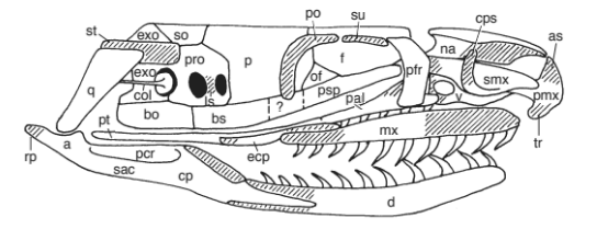
| The right side of the skull of an alethinophidian snake (nose pointing to the right). Bones with teeth are the maxilla (mx), palatine (pal), pterygoid (pt), and dentary (d). From Cundall & Irish 2008. For a key to all abbreviations, click here. The bones or parts of bones that are shaded are not present in all snake species. |
 |
| The right half of the skull of a snake, looking up from the bottom (nose pointing to the right). Bones with teeth are the maxilla (mx), palatine (pal), pterygoid (pt), and dentary (d). The premaxilla (pmx) has no teeth. From Cundall & Irish 2008. For a key to all abbreviations, click here.The bones or parts of bones that are shaded are not present in all snake species. |
 |
| Tooth marks left by a python bite (upper jaw above, lower jaw below). |
 |
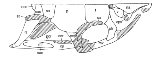
| The right half of the skull of a snake, looking down from the top (nose pointing to the right). No teeth are visible. From Cundall & Irish 2008. For a key to all abbreviations, click here.The bones or parts of bones that are shaded are not present in all snake species. |
 |
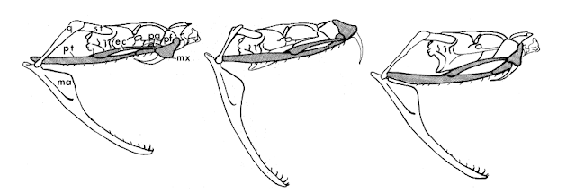
Roughly the same fang movements are made during striking and swallowing. Supratemporal (st), quadrate (q), mandible (ma), pterygoid (pt), ectopterygoid (ec), palatine (pa), prefrontal (pf), maxilla (mx). From Kardong 1977 |
 |
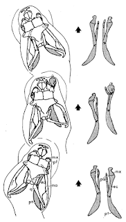
| The independent left and right movement of the upper jaws of a viper. Abbreviations as above. From Kardong 1977. |
The lower jaws or mandible participate in the process of feeding as well, and unlike in humans they have a loose attachment of the lower jaws to each other at the front of the dentary bones. The dentary bones are connected firmly at the back to the articular bones, which are connected to the quadrate bones at a flexible joint, which are connected to the back of the braincase by the supratemporal bones, also at a somewhat moveable joint. Together with the flexible palato-maxillary apparatus ("upper jaws"), this three-part lower jaw allows snakes to open their mouths very wide, walk their heads over, and consume things that are as big as they are without breaking them into smaller pieces or using their non-existent hands. The quadrate also attaches to the columella, which is the sole inner ear bone in reptiles; thus, the lower jaw also conducts sound to the ear.
So there you have it. The snake skull is divided into four functional units: the braincase, the snout, the palato-maxillary apparatus ("upper jaws") and the mandibular apparatus ("lower jaws"), each of which can move independently (well, except for the braincase, which is relatively stationary). The upper jaws are divided into two partially separated structural-functional units, a medial swallowing unit and a lateral prey capture unit, both of which work with the lower jaws to accomplish their tasks.
 |
| From Frazzetta 1970. Click for larger size. |
1 1: Although the pterygoids are stand-alone bones in the roof of the mouth of many vertebrates, in humans they are called the pterygoid processes of the sphenoid bone because they are fused to the sphenoid bone.↩
2 2: I don't necessarily recommend this, partly because if you've been bitten then it's too late, and partly because it's better just to learn the few venomous snake species that live in your area than it is to try to follow some "rule" that inevitably has exceptions.↩
3 Atractaspidids have a ball-and-socket joint between the prefrontal (part of the braincase) and the maxilla, which along with a gap, bridged by a ligament, between the pterygoid and palatine, allows them to "strike" with their fang backwards, with a closed mouth, using just the fang on one side, a useful if terrifying adaptation for envenomating prey in underground burrows. A hook-like ridge on the fang increases the size of the wound, presumably enhancing the absorption of venom.↩
Albright, R. G. and E. M. Nelson. 1959. Cranial kinetics of the generalized colubrid snake Elaphe obsoleta quadrivittata. I. Descriptive morphology. Journal of Morphology 105:193-239.
Albright, R. G. and E. M. Nelson. 1959. Cranial kinetics of the generalized colubrid snake Elaphe obsoleta quadrivittata. II. Functional morphology. Journal of Morphology 105:241-291.
Cundall, D. 1983. Activity of head muscles during feeding by snakes: a comparative study. American Zoologist 23:383-396.
Cundall, D. and H. W. Greene. 2000. Feeding in snakes. Pages 293–333 in K. Schwenk, editor. Feeding: Form, Function, and Evolution in Tetrapod Vertebrates. Academic Press, San Diego, CA.
Cundall, D. and F. Irish. 2008. The snake skull. Pages 349-692 in C. Gans, A. S. Gaunt, and K. Adler, editors. Biology of the Reptilia. Volume 20, Morphology H. The Skull of Lepidosauria. The University Of Chicago Press, Chicago, Illinois, USA <link>
Frazzetta. T. 1970. From hopeful monsters to bolyerine snakes? The American Naturalist 104:55-72 <link>
Frazzetta, T. 1971. Notes upon the jaw musculature of the Bolyerine snake, Casarea dussumieri. Journal of Herpetology 5:61-63
Irish, F. and P. Alberch. 1989. Heterochrony in the evolution of bolyeriid snakes. Fortschritte der Zoolologie 35:205.
Juckett, G. and J. G. Hancox. 2002. Venomous snakebites in the United States: management review and update. American Family Physician 65:1367-1375 <link>
Kardong, K. 1974. Kinesis of the jaw apparatus during the strike in the cottonmouth snake, Agkistrodon piscivorus. Forma et functio 7:327-354.
Kardong, K. V. 1977. Kinesis of the jaw apparatus during swallowing in the cottonmouth snake, Agkistrodon piscivorus. Copeia 1977:338-348 <link>
Lombard, R. E., H. Marx, and G. B. Rabb. 1986. Morphometrics of the ectopterygoid in advanced snakes (Colubroidea): a concordance of shape and phylogeny. Biological Journal of the Linnean Society 27:133-164 <link>
Maisano, J. A. and O. Rieppel. 2007. The skull of the Round Island boa, Casarea dussumieri Schlegel, based on high-resolution X-ray computed tomography. Journal of Morphology 268:371-384 <abstract>
Raynaud, A. 1985. Development of Limbs and Embryonic Limb Reduction. Pages 59-148 in C. Gans and F. Billett, editors. Biology of the Reptilia. Volume 15. Development B. John Wiley & Sons, New York <link>
Rieppel, O. 2012. “Regressed” Macrostomatan Snakes. Fieldiana Life and Earth Sciences 5:99-103 <link>
